Brain chemical sleep
Brain Basics: Understanding Sleep | National Institute of Neurological Disorders and Stroke
Image
Sleep is an important part of your daily routine—you spend about one-third of your time doing it. Quality sleep – and getting enough of it at the right times -- is as essential to survival as food and water. Without sleep you can’t form or maintain the pathways in your brain that let you learn and create new memories, and it’s harder to concentrate and respond quickly.
Sleep is important to a number of brain functions, including how nerve cells (neurons) communicate with each other. In fact, your brain and body stay remarkably active while you sleep. Recent findings suggest that sleep plays a housekeeping role that removes toxins in your brain that build up while you are awake.
Everyone needs sleep, but its biological purpose remains a mystery. Sleep affects almost every type of tissue and system in the body – from the brain, heart, and lungs to metabolism, immune function, mood, and disease resistance. Research shows that a chronic lack of sleep, or getting poor quality sleep, increases the risk of disorders including high blood pressure, cardiovascular disease, diabetes, depression, and obesity.
Sleep is a complex and dynamic process that affects how you function in ways scientists are now beginning to understand. This booklet describes how your need for sleep is regulated and what happens in the brain during sleep.
Anatomy of Sleep
Several structures within the brain are involved with sleep.
Image
The hypothalamus, a peanut-sized structure deep inside the brain, contains groups of nerve cells that act as control centers affecting sleep and arousal. Within the hypothalamus is the suprachiasmatic nucleus (SCN) – clusters of thousands of cells that receive information about light exposure directly from the eyes and control your behavioral rhythm. Some people with damage to the SCN sleep erratically throughout the day because they are not able to match their circadian rhythms with the light-dark cycle. Most blind people maintain some ability to sense light and are able to modify their sleep/wake cycle.
The brain stem, at the base of the brain, communicates with the hypothalamus to control the transitions between wake and sleep. (The brain stem includes structures called the pons, medulla, and midbrain.) Sleep-promoting cells within the hypothalamus and the brain stem produce a brain chemical called GABA, which acts to reduce the activity of arousal centers in the hypothalamus and the brain stem. The brain stem (especially the pons and medulla) also plays a special role in REM sleep; it sends signals to relax muscles essential for body posture and limb movements, so that we don’t act out our dreams.
The thalamus acts as a relay for information from the senses to the cerebral cortex (the covering of the brain that interprets and processes information from short- to long-term memory). During most stages of sleep, the thalamus becomes quiet, letting you tune out the external world.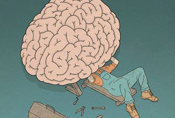 But during REM sleep, the thalamus is active, sending the cortex images, sounds, and other sensations that fill our dreams.
The pineal gland, located within the brain’s two hemispheres, receives signals from the SCN and increases production of the hormone melatonin, which helps put you to sleep once the lights go down. People who have lost their sight and cannot coordinate their natural wake-sleep cycle using natural light can stabilize their sleep patterns by taking small amounts of melatonin at the same time each day. Scientists believe that peaks and valleys of melatonin over time are important for matching the body’s circadian rhythm to the external cycle of light and darkness.
The basal forebrain, near the front and bottom of the brain, also promotes sleep and wakefulness, while part of the midbrain acts as an arousal system. Release of adenosine (a chemical by-product of cellular energy consumption) from cells in the basal forebrain and probably other regions supports your sleep drive.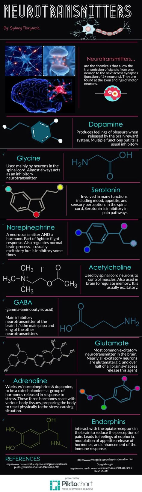 Caffeine counteracts sleepiness by blocking the actions of adenosine.
The amygdala, an almond-shaped structure involved in processing emotions, becomes increasingly active during REM sleep.
Sleep Stages and Mechanisms
Sleep Stages
There are two basic types of sleep: rapid eye movement (REM) sleep and non-REM sleep (which has three different stages). Each is linked to specific brain waves and neuronal activity. You cycle through all stages of non-REM and REM sleep several times during a typical night, with increasingly longer, deeper REM periods occurring toward morning.
Image
Stage 1 non-REM sleep is the changeover from wakefulness to sleep. During this short period (lasting several minutes) of relatively light sleep, your heartbeat, breathing, and eye movements slow, and your muscles relax with occasional twitches. Your brain waves begin to slow from their daytime wakefulness patterns.
Image
Stage 2 non-REM sleep is a period of light sleep before you enter deeper sleep. Your heartbeat and breathing slow, and muscles relax even further. Your body temperature drops and eye movements stop. Brain wave activity slows but is marked by brief bursts of electrical activity. You spend more of your repeated sleep cycles in stage 2 sleep than in other sleep stages.
Image
Stage 3 non-REM sleep is the period of deep sleep that you need to feel refreshed in the morning. It occurs in longer periods during the first half of the night. Your heartbeat and breathing slow to their lowest levels during sleep. Your muscles are relaxed and it may be difficult to awaken you. Brain waves become even slower.
Image
REM sleep first occurs about 90 minutes after falling asleep. Your eyes move rapidly from side to side behind closed eyelids. Mixed frequency brain wave activity becomes closer to that seen in wakefulness. Your breathing becomes faster and irregular, and your heart rate and blood pressure increase to near waking levels. Most of your dreaming occurs during REM sleep, although some can also occur in non-REM sleep. Your arm and leg muscles become temporarily paralyzed, which prevents you from acting out your dreams. As you age, you sleep less of your time in REM sleep. Memory consolidation most likely requires both non-REM and REM sleep.
Sleep Mechanisms
Two internal biological mechanisms–circadian rhythm and homeostasis–work together to regulate when you are awake and sleep.
Circadian rhythms direct a wide variety of functions from daily fluctuations in wakefulness to body temperature, metabolism, and the release of hormones. They control your timing of sleep and cause you to be sleepy at night and your tendency to wake in the morning without an alarm. Your body’s biological clock, which is based on a roughly 24-hour day, controls most circadian rhythms. Circadian rhythms synchronize with environmental cues (light, temperature) about the actual time of day, but they continue even in the absence of cues.
Image
Your body's biological clock is based on a 24-hour day and controls most circadian rhythms. These rhythms affect a variety of functions including body temperature (represented as the white line on the chart above). Melatonin - a hormone released by the pineal gland - helps you feel sleepy once the lights go down. The peaks and valleys of melatonin (represented as the gold line above) are important for matching the body's circadian rhythm to the external cycle of light and darkness.Sleep-wake homeostasis keeps track of your need for sleep. The homeostatic sleep drive reminds the body to sleep after a certain time and regulates sleep intensity. This sleep drive gets stronger every hour you are awake and causes you to sleep longer and more deeply after a period of sleep deprivation.
Factors that influence your sleep-wake needs include medical conditions, medications, stress, sleep environment, and what you eat and drink. Perhaps the greatest influence is the exposure to light. Specialized cells in the retinas of your eyes process light and tell the brain whether it is day or night and can advance or delay our sleep-wake cycle. Exposure to light can make it difficult to fall asleep and return to sleep when awakened.
Night shift workers often have trouble falling asleep when they go to bed, and also have trouble staying awake at work because their natural circadian rhythm and sleep-wake cycle is disrupted. In the case of jet lag, circadian rhythms become out of sync with the time of day when people fly to a different time zone, creating a mismatch between their internal clock and the actual clock.
How Much Sleep Do You Need?
Image
Sleep needs change with age as shown on the chart above. Initially, babies sleep 16-18 hours a day. School-age children and teens need about 9.5 hours of sleep each night. Most adults require 7-9 hours of sleep at night. However, older adults (age 60 and above) tend to sleep for shorter periods at night.Your need for sleep and your sleep patterns change as you age, but this varies significantly across individuals of the same age. There is no magic “number of sleep hours” that works for everybody of the same age. Babies initially sleep as much as 16 to 18 hours per day, which may boost growth and development (especially of the brain). School-age children and teens on average need about 9.5 hours of sleep per night. Most adults need 7-9 hours of sleep a night, but after age 60, nighttime sleep tends to be shorter, lighter, and interrupted by multiple awakenings. Older people are also more likely to take medications that interfere with sleep.
In general, people are getting less sleep than they need due to longer work hours and the availability of round-the-clock entertainment and other activities.
Many people feel they can "catch up" on missed sleep during the weekend but, depending on how sleep-deprived they are, sleeping longer on the weekends may not be adequate.
Dreaming and Sleep Tracking
Dreaming
Everyone dreams. You spend about 2 hours each night dreaming but may not remember most of your dreams. Its exact purpose isn’t known, but dreaming may help you process your emotions. Events from the day often invade your thoughts during sleep, and people suffering from stress or anxiety are more likely to have frightening dreams. Dreams can be experienced in all stages of sleep but usually are most vivid in REM sleep. Some people dream in color, while others only recall dreams in black and white.
Image
Tracking Sleep Through Smart Technology
Millions of people are using smartphone apps, bedside monitors, and wearable items (including bracelets, smart watches, and headbands) to informally collect and analyze data about their sleep. Smart technology can record sounds and movement during sleep, journal hours slept, and monitor heart beat and respiration. Using a companion app, data from some devices can be synced to a smartphone or tablet, or uploaded to a PC. Other apps and devices make white noise, produce light that stimulates melatonin production, and use gentle vibrations to help us sleep and wake.
The Role of Genes and Neurotransmitters
Chemical signals to sleep
Clusters of sleep-promoting neurons in many parts of the brain become more active as we get ready for bed. Nerve-signaling chemicals called neurotransmitters can “switch off” or dampen the activity of cells that signal arousal or relaxation. GABA is associated with sleep, muscle relaxation, and sedation. Norepinephrine and orexin (also called hypocretin) keep some parts of the brain active while we are awake. Other neurotransmitters that shape sleep and wakefulness include acetylcholine, histamine, adrenaline, cortisol, and serotonin.
Genes and sleep
Genes may play a significant role in how much sleep we need. Scientists have identified several genes involved with sleep and sleep disorders, including genes that control the excitability of neurons, and "clock" genes such as Per, tim, and Cry that influence our circadian rhythms and the timing of sleep.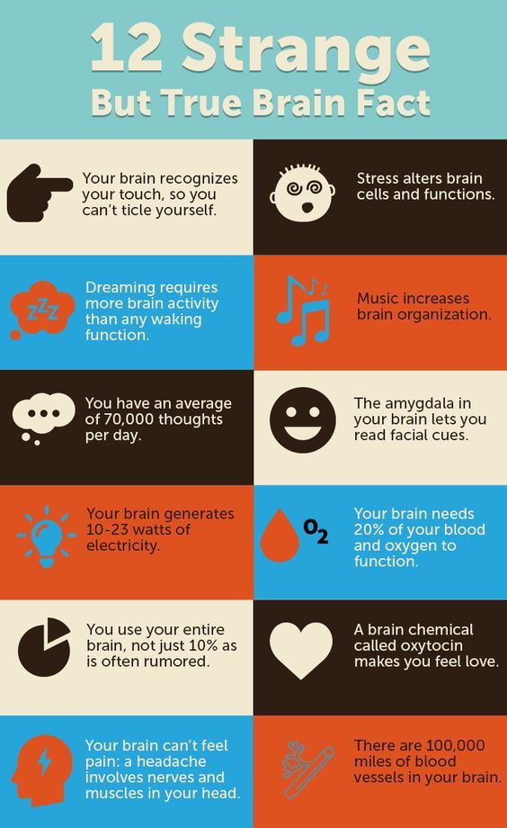 Genome-wide association studies have identified sites on various chromosomes that increase our susceptibility to sleep disorders. Also, different genes have been identified with such sleep disorders as familial advanced sleep-phase disorder, narcolepsy, and restless legs syndrome. Some of the genes expressed in the cerebral cortex and other brain areas change their level of expression between sleep and wake. Several genetic models–including the worm, fruit fly, and zebrafish–are helping scientists to identify molecular mechanisms and genetic variants involved in normal sleep and sleep disorders. Additional research will provide better understand of inherited sleep patterns and risks of circadian and sleep disorders.
Image
Sleep studies
Your health care provider may recommend a polysomnogram or other test to diagnose a sleep disorder. A polysomnogram typically involves spending the night at a sleep lab or sleep center. It records your breathing, oxygen levels, eye and limb movements, heart rate, and brain waves throughout the night. Your sleep is also video and audio recorded. The data can help a sleep specialist determine if you are reaching and proceeding properly through the various sleep stages. Results may be used to develop a treatment plan or determine if further tests are needed.
Tips for Getting a Good Night's Sleep
Image
Getting enough sleep is good for your health. Here are a few tips to improve your sleep:
- Set a schedule – go to bed and wake up at the same time each day.
- Exercise 20 to 30 minutes a day but no later than a few hours before going to bed.
- Avoid caffeine and nicotine late in the day and alcoholic drinks before bed.
- Relax before bed – try a warm bath, reading, or another relaxing routine.
- Create a room for sleep – avoid bright lights and loud sounds, keep the room at a comfortable temperature, and don’t watch TV or have a computer in your bedroom.
- Don’t lie in bed awake. If you can’t get to sleep, do something else, like reading or listening to music, until you feel tired.
- See a doctor if you have a problem sleeping or if you feel unusually tired during the day. Most sleep disorders can be treated effectively.
Hope Through Research
Scientists continue to learn about the function and regulation of sleep. A key focus of research is to understand the risks involved with being chronically sleep deprived and the relationship between sleep and disease. People who are chronically sleep deprived are more likely to be overweight, have strokes and cardiovascular disease, infections, and certain types of cancer than those who get enough sleep. Sleep disturbances are common among people with age-related neurological disorders such as Alzheimer’s disease and Parkinson’s disease. Many mysteries remain about the association between sleep and these health problems. Does the lack of sleep lead to certain disorders, or do certain diseases cause a lack of sleep? These, and many other questions about sleep, represent the frontier of sleep research.
Sleep/Wake Cycles | Johns Hopkins Medicine
Sometimes you drift off to sleep easily. Other times you toss and turn for hours before you slip into a fitful sleep. After lunch you may be dragging. Later, your energy levels soar just in time for bed.
How and when you feel sleepy has to do with your sleep/wake cycles. These cycles are triggered by chemicals in the brain.
Brain chemicals and sleep
Chemicals called neurotransmitters send messages to different nerve cells in the brain. Nerve cells in the brainstem release neurotransmitters. These include norepinephrine, histamine, and serotonin. Neurotransmitters act on parts of the brain to keep it alert and working well while you are awake.
Other nerve cells stop the messages that tell you to stay awake. This causes you to feel sleepy. One chemical involved in that process is called adenosine. Caffeine promotes wakefulness by blocking the receptors to adenosine. Adenosine seems to work by slowly building up in your blood when you are awake. This makes you drowsy. While you sleep, the chemical slowly dissipates.
Sleep processes
Two body processes control sleeping and waking periods. These are called sleep/wake homeostasis and the circadian biological clock.
With sleep/wake homeostasis, the longer you are awake, the greater your body senses the need to sleep. If this process alone was in control of your sleep/wake cycles, in theory you would have the most energy when you woke up in the morning. And you would be tired and ready for sleep at the end of the day.
But your circadian biological clock causes highs and lows of sleepiness and wakefulness throughout the day. Typically, most adults feel the sleepiest between 2 a.m. and 4 a.m., and also between 1 p.m. and 3 p.m. Getting plenty of regular sleep each night can help to balance out these sleepy lows.
Your body’s internal clock is controlled by an area of the brain called the SCN (suprachiasmatic nucleus). The SCN is located in the hypothalamus.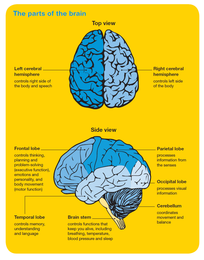 The SCN is sensitive to signals of dark and light. The optic nerve in your eyes senses the morning light. Then the SCN triggers the release of cortisol and other hormones to help you wake up. But when darkness comes at night, the SCN sends messages to the pineal gland. This gland triggers the release of the chemical melatonin. Melatonin makes you feel sleepy and ready for bed.
Neurotransmitters and your sleep
Some neurotransmitters help your body recharge while you sleep. They can even help you to remember things that you learned, heard, or saw while you were awake. The neurotransmitter acetylcholine is at its strongest both during REM (rapid eye movement) sleep and while you are awake. It seems to help your brain keep information gathered while you are awake. It then sets that information as you sleep. So if you study or learn new information in the hours before bed, "sleeping on it" can help you remember it.
Other neurotransmitters may work against you as you sleep. Abnormalities with the neurotransmitter dopamine may trigger sleep disorders such as restless legs syndrome.
Even losing just 1 hour of sleep over a few days can have an effect. It can lead to a decrease in performance, mood, and thinking. Getting regular, adequate amounts of sleep is important. It can help you feel awake and refreshed during the day. It can also help you feel relaxed and sleepy at night. This helps make you ready for a long, restful night of sleep.
What does our brain do while we sleep
While you sleep, the brain works. One of the reasons why you should not neglect sleep is that during the sleep period your brain processes all the information received during the day. He puts it on the shelves and makes it so that unnecessary data is eliminated, and important data is fixed in memory. We will tell you what our brain does during sleep and how it is related to learning.
What happens to us in sleep
In 1928, the German scientist Hans Berger first recorded the electrical activity of the human brain - an electroencephalogram (EEG). So, he proved that the cerebral cortex is busy with activities. In addition, scientists observed waves with high-amplitude oscillations with a frequency of 0.5-4 Hz, which they later called slow, and the sleep phase became slow-wave sleep.
Slow consists of four stages:
-
The first stage is between wakefulness and falling asleep;
-
On the second, “light” sleep is noted, when the pulse rate and breathing are regulated, the body temperature drops;
-
The third and fourth stages are deep sleep.
REM sleep accounts for 20-25% of a night's sleep, one phase lasts 10-20 minutes and alternates with non-REM sleep. So, the cycle is repeated on average 4-5 times. It used to be that REM sleep was important for learning and memory. However, new research suggests that non-REM sleep is more important for strengthening memory and processing the information received. Also, this phase is more relaxed.
Parts of the brain and their functions during sleep
The hypothalamus contains groups of nerve cells that act as control centers that influence sleep and arousal. Inside the hypothalamus is the suprachiasmatic nucleus (SCN), which receives information about light exposure.
The brainstem (comprises structures called the pons, medulla, and midbrain) controls the transitions between wakefulness and sleep. A chemical (GABA) is produced in the hypothalamus and brain stem, which reduces the activity of the excitation centers. The brain stem sends signals to relax the muscles.
The thalamus transmits information from the senses to the cerebral cortex. It is active during REM sleep and sends images, sounds and other sensations that are in dreams to the cerebral cortex.
The pineal gland receives signals from the suprachiasmatic nucleus (SCN) in the hypothalamus and increases the production of the hormone melatonin. It is what promotes sleep.
The basal forebrain promotes sleep and wakefulness, while part of the midbrain, on the contrary, excites. When adenosine is released from the basal forebrain, the urge to sleep appears.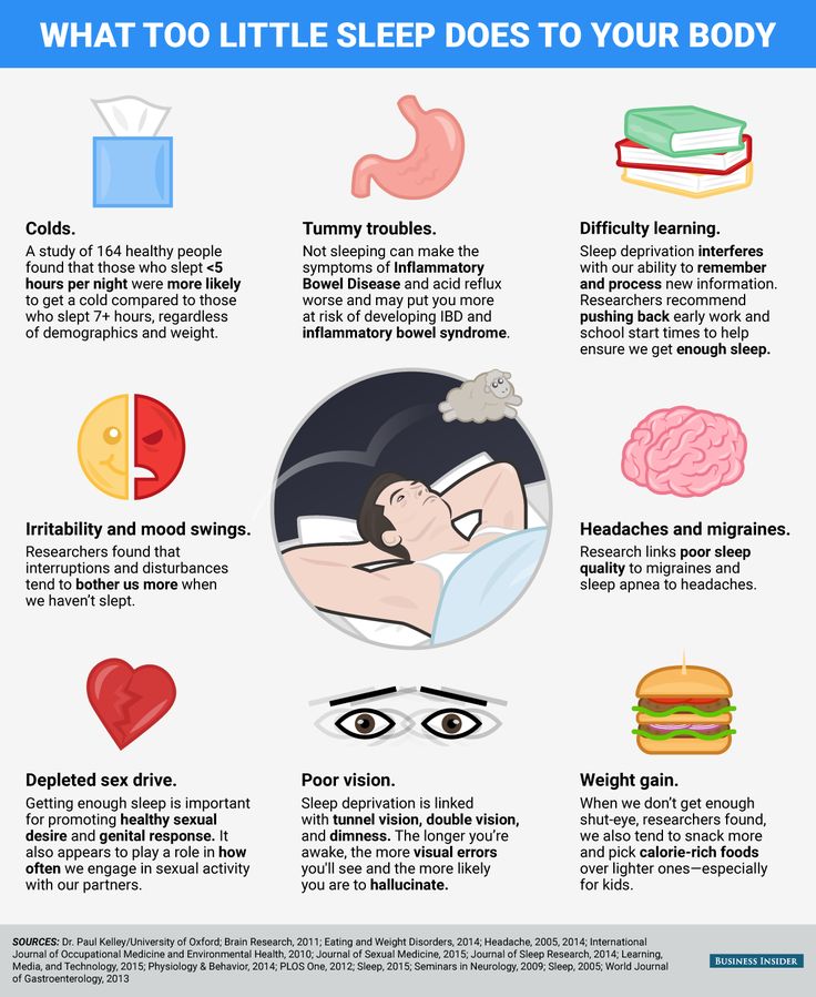
The amygdala becomes more active during REM sleep. It is responsible for regulating emotions.
How healthy sleep helps you learn more effectively
Multiple hypotheses explain the links between sleep and learning in humans. Research shows that sleep allows not only rest, but also information processing.
REM sleep and NREM sleep play different roles in memory consolidation. REM is associated with the consolidation of non-declarative memories. For example, these are automatic actions such as riding a bike, brushing your teeth. Non-REM sleep (NREM) is associated with the consolidation of declarative memories. These are facts that are not simply remembered: for example, historical dates, new terms. For example, sleep helps the brain edit memory according to the synaptic scaling hypothesis.
Sleep provides more efficient and effective storage of information needed for learning
According to a study by UC Berkeley professor of neuroscience and psychology Matthew Walker, it was found that sleep fixes declarative memories. In the study, two groups learned word pairs and then either fell asleep or didn't sleep and were tested again. The other two groups did the same, except that they also learned other pairs of words right before the retest to try and make it harder to remember previously learned words. But thanks to sleep, they remained in memory.
Sleep helps people re-examine their memories. The same patterns of brain activity that occur during learning have been found to repeat during sleep, only faster. Sleep compensates for the strengthening of some synapses (connections) between neurons due to weakening. This allows other connections to be strengthened while we are awake.
Learning is a process of strengthening neural connections
In learning, such a process as consolidation is important - the transition from slow conscious information processing to fast and unconscious automation. So, if you know how to perform some actions without much effort, there is an opportunity to improve the skill or get a new one. Sleep helps consolidation. Thus, the brain launches a special algorithm that reproduces the events of the past day and transfers it to the compartment of our memory, writes Stanislas Dean, author of How We Learn.
“In 1994, Israeli scientists conducted an experiment that confirmed this. “Over the course of the day, the volunteers learned to detect the band at a specific point on the retina. Task performance slowly increased until it reached a plateau. However, after sleep, the performance of the participants increased dramatically and remained at this level for the next few days. When the researchers woke the subjects during REM sleep, there was no improvement. Therefore, it is non-REM sleep that promotes consolidation, while REM sleep promotes the consolidation of perceptual and motor skills.”
Another study based on electrophysiological recordings of the prefrontal cortex of rats found that clusters of cells that formed during learning were more active during subsequent sleep episodes. These reproductions were more prominent during non-REM sleep and were accompanied by hippocampal reactivation. Thus, neural patterns in large brain networks are marked during learning, so they are reproduced and presumably consolidated during subsequent sleep.
Lyubov Karas
Tags
#self-development
#well-being
#erudition
Progress in the study of sleep in the era of electrophysiology. Visceral Theory of Sleep
In the previous issue of this journal, devoted to the problems of somnology [1], a number of forgotten, but important, from our point of view, works carried out before the era of electrophysiology were presented to the attention of readers. That review was intended as an introduction to prepare readers for a new approach to the study of sleep, which led to the emergence of a theory explaining the purpose of this bodily state. This article can also be read as an independent work, but the history of approaching the understanding of the functional purpose of sleep will be more complete if you first read the contents of the previous short publication.
It should be noted that this review is not strictly historical. Regarding the development of sleep research, we provide a minimum of references, since most of the referenced works are well known and have been widely presented. You can, for example, refer to the monograph by V.M. Kovalzon [2], where there are necessary bibliographic references. This publication highlights lesser-known works and studies that have made a special contribution to understanding the functional purpose of sleep.
The contribution of electrophysiology methods to changing ideas about the essence of sleep
The previous article [1] ended with a description of the experiments carried out in the laboratory of acad. K.M. Bykov in Leningrad in 1936. As in much earlier experiments by M.M. Manaseina, the study showed that sleep deprivation leads to numerous pathological abnormalities in the work of visceral systems and the inevitable death of animals. When these behavioral experiments were carried out, the era of electrophysiology was already beginning in somnology, opening up new horizons in the study of the functioning of the brain and, in particular, in the study of sleep.
Shortly after the discovery by H. Berger [3] of the method of electroencephalography and the recording of the first electroencephalogram (EEG), A. Lumis et al. [4, 5] in the United States studied in detail the electrical activity of the human brain in the state of sleep and described the dynamics of the appearance of sleep spindles, K-complexes, and slow-wave activity during the transition from wakefulness to sleep. A year after the publication of their work, the German scientist R. Klaue [6] recorded an EEG from the surface of the dura mater and directly from the gray matter of the cerebral cortex of a naturally sleeping cat. It has been shown that during the transition from wakefulness to sleep, the same changes occur in the electrical activity of the cat's brain as in the human EEG. But in the experiments of R. Klaue, a new phenomenon was also discovered - the disappearance of slow waves (EEG desynchronization) during periods of the deepest sleep. Now this state is called the phase of REM, or paradoxical, sleep. In foreign literature, the term "REM sleep" is adopted to denote this state ( Rapid eye movement sleep). As often happens in science, the discovery of REM sleep is unfairly associated with the names of K. Aserinsky and N. Kleitman [7], who noticed and described this phenomenon in humans 20 years after the publication of R. Klaue's work [6].
Pronounced changes in the nature of the electrical activity of the brain during the transition from quiet wakefulness to sleep made the EEG an integral component of the study of sleep for many years. At the same time, this led to an erroneous judgment that the EEG pattern is an objective indicator of the onset of sleep. The researchers forgot that the primary ones were still observations of the behavioral criteria of the upcoming sleep, and EEG changes only correlated well in time with these moments.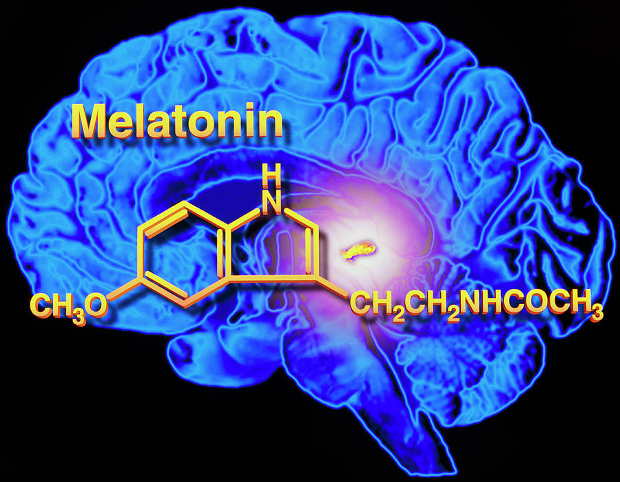
The observed striking changes in the structure of the EEG as sleep progressed supported the conviction of most scientists that the state of sleep is primarily important for the functioning of the brain. The appearance of slow waves in the EEG was considered as a reflection of a special state of the brain during sleep, the functional purpose of which remained a mystery. The attention of somnologists was again focused on the study of brain processes during periods of sleep, and the previously discovered dramatic visceral disorders accompanying sleep deprivation again faded into the background and were ignored for a long time when discussing the functional purpose of this state.
The new era of electrophysiology was not limited to EEG recording. The most important for somnology were the results of experiments with recording the activity of single neurons of the cerebral cortex during the development of sleep. It was found that during sleep, the average frequency of impulse activity of cortical neurons not only did not decrease, but could even increase.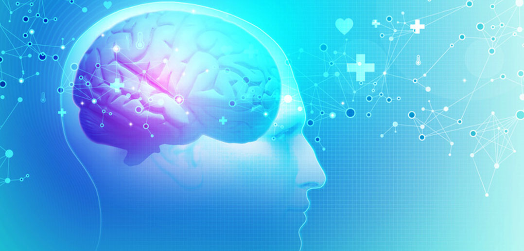 Indeed, during periods of slow sleep, the character of impulse discharges of cortical neurons changed. Even with the preservation of the average frequency of impulses, the neurons changed the structure of impulse discharges. The pulses were grouped into high-frequency bursts, separated by pauses of silence. The rhythm of bursts of neuronal activity often coincided well with the rhythm of slow EEG waves. The activation of neurons became especially strong in the phase of REM (paradoxical) sleep, when individual high-frequency bursts of impulses merged into an almost continuous high-frequency discharge, which continued until the end of this phase of sleep.
The results of these observations, repeated many times in different laboratories, undoubtedly demanded that the idea of sleep be abandoned as a diffuse cortical inhibition. The nature of neuronal activity during sleep made it possible to draw another very important conclusion. It became clear that the activity of cortical neurons does not have a rigid and unambiguous connection with the behavior of the animal. Thus, the neurons of the visual cortex in wakefulness responded with impulse discharges to the appearance and movement of objects in the visual field. The activation of neurons in the motor or somatosensory cortex was associated with certain movements of the animal. And during sleep, similar activity of the same neurons in the visual or somatosensory cortex occurred in the complete absence of any visual stimuli or motor activity. At the same time, the thresholds for sensory perception during sleep rose so much that real visual or somatic stimuli did not lead to the activation of these neurons.
After I.P. Pavlova's realization of higher mental, or, as they are more commonly called now, cognitive functions, was associated with the work of the cerebral cortex. The nature of the activity of neurons in the cerebral cortex during sleep definitely contradicted this conclusion. Obviously, during sleep, cognitive functions fade or are minimized. And the activity of neurons in all cortical zones during sleep remains at the same level or even increases.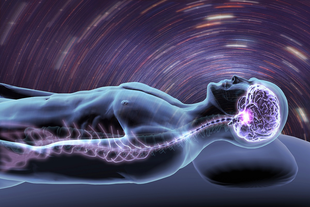 The dream remained an incomprehensible phenomenon. No wonder the motto of one of the last congresses of the World Association of Sleep Researchers was "The Mystery of Sleep" ("mystery of sleep").
Simultaneously with the study of EEG features and neuronal activity in the sleep-wake cycle, the method of microelectrostimulation played an important role in the study of brain structures involved in the transition from wakefulness to sleep. In the brainstem, from the pons to the hypothalamus, a dozen and a half small loci have been described, local stimulation of which caused the development of non-REM sleep, REM sleep, or awakening [8–10]. Despite the complexity and ambiguity of the interpretation of the effects of electrical stimulation, an anatomical picture of the system of brain structures, to one degree or another involved in sleep control, began to take shape. It became obvious that there was no single center that controlled the sleep-wake cycle. However, the main question remained unanswered: by what signal and for what purpose did the neurons begin to activate, leading to falling asleep and, accordingly, why did the activation of other neurons suddenly occur, returning the body to the state of wakefulness. But the advances in electrophysiology gave reason to think that answers to these questions could soon be obtained.
Limitations in electroencephalographic sleep studies
As applied to humans, the electroencephalography method until recently had one serious limitation. Head and body movements resulted in strong artifacts due to the movement of numerous wires and the resulting microdisplacements of the electrodes. But during the period of restful sleep, and especially in the phase of REM sleep with complete atony of the skeletal muscles, there were no movements and nothing interfered with obtaining a clear signal. But pure encephalographic signals during the period of wakefulness could only be obtained under unnatural conditions of the most immobile state and low levels of illumination and noise, which are common for somnological laboratories.
After the start of EEG studies in animals with skull-implanted recording electrodes and amplifiers mounted directly on the animal's head, the problem of movement artifacts was solved. At the same time, it turned out that the EEG pattern in wakefulness is not so unambiguous. During active and rhythmic movements of the extremities, slow waves appeared in the EEG of the somatosensory areas of the cortex, often indistinguishable in their characteristics from the waves of slow wave sleep. The use of rhythmic visual stimulation also led to the appearance of slow waves in the visual cortex. In animals, when registering neurons in different areas of the brain, it was possible to obtain a typical "burst activity of sleep" in wakefulness, simply by selecting the appropriate parameters of external stimulation. Here it is necessary to recall that even in the very first studies of the EEG in humans, the reaction of assimilation of the rhythm of flickering light was discovered - in the state of active wakefulness, waves appeared on the EEG with the frequency of flickering of the light stimulus. Gradually, the suspicion grew that the EEG pattern is determined not so much by the state of sleep or wakefulness, but by the nature of the afferent inflow entering the cerebral cortex in a particular state of the animal.
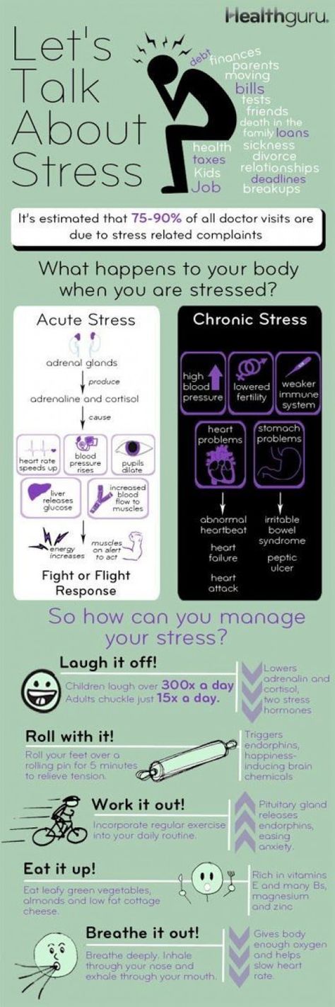
But what kind of afferent inflow in the state of sleep could one speak of? It was generally accepted that during sleep, the flow of sensory information from the outside world and from the proprioceptors of the body to the cerebral cortex is very difficult. Firstly, purely mechanical barriers worked - dark, quiet and soft places were chosen for sleep, and the eyes were additionally closed for centuries. But, in addition, additional neural mechanisms for blocking the transmission of sensory information to the corresponding areas of the cerebral cortex were also described [11]. The cerebral cortex was considered to be completely isolated from the afferent inflow during sleep. But was this representation logically flawless? Within the framework of the classical neurophysiological paradigm of the middle of the 20th century, probably yes!
Nevertheless, at that time science was entering a new - informational - era. The foundations of information theory were laid, the first computers based on universal processors were created. It became obvious that it was general-purpose, and not specialized, processors that would determine the future of the information age. The work of computer processors in the shared time mode, when the same processor was involved in the work for "different users", became common. These achievements of the informational approach could not but be reflected in the views of neurophysiologists on the structure and functions of the most advanced computer, which is the brain.
Within the framework of these ideas, nothing ruled out the possibility of using the same brain structure to analyze one stream of information in the waking state and another in the dream state.
But what information could the brain process during sleep? The answer to this question seemed to be the most important for understanding the functional purpose of this state. And it seemed that the answer to this question lay on the surface. It was enough to recall the results of experiments in which animals without sleep soon died from deviations in the visceral sphere. These old observations were supported by a series of new works carried out as early as 90s in the laboratory of one of the leading experts in human sleep physiology, A. Rechtshaffen [12]. Experiments conducted on rats were devoted to the study of the consequences of complete sleep deprivation. For this purpose, a new and original technique was developed, which made it possible to convincingly separate the effect of sleep deprivation from the possible negative effect of stress associated with the procedure for maintaining uninterrupted wakefulness. For the first time, computer analysis of the EEG was used, which made it possible to automatically determine the beginning of falling asleep and turn on the corresponding system for awakening the animal. Not knowing about the research of M.M. Manaseina (end of the 19th century) and the work of the laboratory staff of K.M. Bykov (beginning of the 20th century), similar results were obtained in the laboratory of A. Rechtshaffen at a new technical level.
 Sleep deprivation inevitably led to the death of animals from a variety of pathological abnormalities in almost all visceral systems of the body. Surprisingly, no traces of degeneration were found in the brains of animals that died from deprivation [13].
Visceral hypothesis of the functional purpose of sleep and the results of experiments to test its non-trivial predictions
The answer to the key question of the physiology of sleep suggests itself. But what if during sleep the brain simply switches from analyzing exteroceptive information coming from the outside world and proprioceptive information about the position of one’s own body to analyzing interoceptive information from all life support systems of the body in order to diagnose their condition and develop ways to restore the efficiency of all these systems? What if it is the rhythmic flow of signals from the peristaltic organs of the gastrointestinal tract, from the rhythmic contractions of the heart and lungs that determines the wave character of the EEG in the non-REM sleep phase? And information from organs that do not have a pronounced rhythm, such as the liver, kidneys, organs of the reproductive system, and, finally, the brain itself, which is also a visceral organ, can be processed during REM sleep? Thus, in one complete sleep cycle, the brain could scan the state of all life support systems.
The proposed hypothesis for the first time made it possible to explain by one assumption all the known phenomenology associated with the state of sleep and made understandable the reasons for the dramatic consequences of its deprivation. An important element of this hypothesis was the fact that the nontrivial predictions that followed from it could be verified in simple experiments.
The first such key prediction was the expected switching of neurons in the sensory areas of the cerebral cortex to the analysis of interoceptive information in the sleep state. It was from this that the cycle of works on testing the stated hypothesis was started.
Primary visual cortex of cats was chosen for experiments. The choice of this zone was determined by the fact that at that time this zone of the cortex was the most thoroughly studied, and no one doubted that the neurons of this zone of the cerebral cortex are involved exclusively in the analysis of visual information. In addition, one of the authors of this article (I.P.) had extensive experience with neurons in the visual cortex of cats in chronic experiments.
In awake cats in the primary visual cortex (field 17), the impulse activity of a group of neurons responding to visual stimulation was recorded. When the cat fell asleep, electrical stimulation of the intestines was carried out, paying special attention to the fact that the stimulating current did not wake the sleeping animal. We soon saw that intestinal stimulation not only did not wake the cat, but, on the contrary, deepened sleep. But, of course, the main result was not the deepening of sleep as a result of electrical stimulation, but the fact that the “visual” neurons actually began to respond to intestinal stimulation during sleep. After awakening, these responses disappeared, and the neurons again became classical visual neurons [14].
It makes no sense to give the technical details of this and all subsequent experiments, since the studies are published and all points of interest can be easily found in the relevant articles.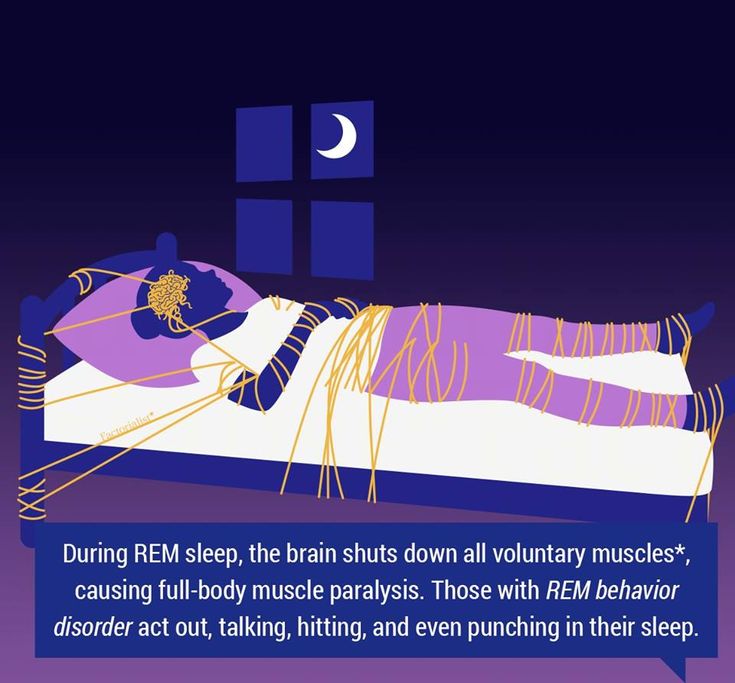 Let us only briefly dwell on the logic of subsequent experiments and on the results obtained.
In order to make sure that the responses of visual cortex neurons to visceral stimulation were not a specific feature of the organism of cats, we carried out similar studies on rabbits and monkeys, recording neuronal impulse activity or using the method of averaging evoked responses in EEG signals [15, 16] . In one study on monkeys, instead of invasive intestinal electrical stimulation, we used non-invasive magnetic stimulation of the abdomen and obtained similar results [17]. The effectiveness of magnetic stimulation opened up the potential for similar studies involving humans.
Opponents of the visceral sleep hypothesis often said that the effects observed in our experiments could reflect known phenomena such as sensory provoked K-complexes. Already in the first encephalographic studies of sleep [4, 5], it was noted that in the sleepy state, various sensory stimuli (auditory or somatic) could cause the appearance of K-complexes in the EEG, typical EEG graph elements in the state of drowsiness, associated with a certain activation of cortical neurons.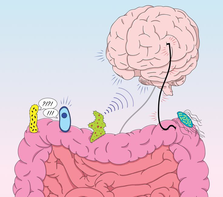 It was assumed that the observed responses of neurons to visceral stimuli could have the same "non-specific" nature. This question has been studied in experiments on rabbits, the results of which showed that sensory provoked K-complexes and cortical responses to visceral stimulation are different physiological phenomena that manifest themselves in different phases of the sleep-wake cycle. Moreover, the study led to the conclusion that sensory-provoked K-complexes most likely reflect bursts of visceral afferentation that breaks through into the cerebral cortex during transitional periods from wakefulness to sleep [18].
However, the first works using electrical or magnetic stimulation of the organs of the gastrointestinal tract, indeed, had one significant drawback. The electrical and magnetic stimuli used in them were artificial. It was important to make sure that the connection of the visceral systems with the neurons of the cerebral cortex is also manifested in the natural conditions of the organism's vital activity.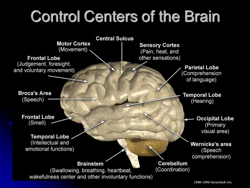 We were able to conduct such a study with the help of our colleagues from the Institute of Physiology. I.P. Pavlov in St. Petersburg prof. V.A. Bagaev (unfortunately, now deceased) and his colleague I.I. Busygina. Cats were implanted with electrodes in the walls of the stomach and duodenum, allowing to record the natural myoelectric activity of these organs. In addition, fistulas were implanted into the wall of the stomach, making it possible to change the intragastric environment.
In a new series of experiments on this methodological basis, it was demonstrated that the natural myoelectric activity of the stomach and intestines in slow-wave sleep is also reflected in the EEG and impulse activity of cortical neurons [19]. It was also shown that a change in the intragastric environment during sleep leads to a sharp change in the pattern of normal slow-wave activity of the cortex and affects the statistical characteristics of the impulse activity of cortical neurons [20]. At the same time, the nature of interneuronal interaction in the cerebral cortex remained practically unchanged during the transition from wakefulness to sleep [21]. This observation testified in favor of the fact that both in wakefulness and in sleep the cerebral cortex carries out similar processing of incoming information. Only the sources of these information flows and the consumers of the results of their processing differ.
Visceral Theory of Sleep
A large number of experiments confirming the hypothesis described above and its ability to explain the entire phenomenology of the state of sleep and suggest probable mechanisms for the appearance of pathological deviations associated with disturbances in the regulation of the sleep-wake cycle, made it possible to speak of this hypothesis as a visceral theory of sleep. On fig. 1 and 2 Fig. 1. Scheme of the main information flows of the body in the waking state. The solid lines are the paths along which the information is transmitted, the dotted line is the inactivated paths. Explanations in the text.Fig. 2. Scheme of the main information flows of the body in a state of sleep. The solid lines are the paths along which the information is transmitted, the dotted line is the inactivated paths. Explanations in the text. the main information flows of the organism in the state of wakefulness and sleep are schematically depicted as they are presented within the framework of this theory.
The most important elements of this system are devices that, depending on the control signal, can close or open the conduction along the nerve pathway. It is natural to assume that this function is performed by synaptic switches. In physiology, for example, the mechanism of presynaptic inhibition or triadic synapses are well known, which are quite suitable for this role. Structures that open or close conduction along a particular pathway are indicated by circles in the figures. Small gray arrows next to the circles indicate incoming signals that control the processes of their switching. A discussion of the nature of these signals will be given below. The white rectangles inside the circles and oriented along the conducting path mean that the conduction in this direction is open. If the rectangles are black and rotated across the direction, then the conduction through this channel is blocked.
In wakefulness (see Fig. 1), information about the environment and the position of the animal's body, perceived by exteroreceptors and proprioreceptors, passes through an open channel through the thalamus and is sent to the cerebral cortex for further processing. The results of this processing are transmitted via open output channels to the block of behavior organization and to the block associated with consciousness. It was said above that it is the study of the activity of cortical neurons in the sleep-wake cycle that leads to the need to separate the structures associated with consciousness from the cortex. This issue is argued in detail in our work [22], which is specially devoted to this issue.
Visceral organs in wakefulness work under the control of the autonomic nervous system. Of course, the individual functions of the visceral organs must be linked to the needs of the current behavior.![]() For example, change the heart rate and respiration rate with a change in physical activity, evaluate the quality and edibility of food at the entrance of the gastrointestinal tract, determine the time and place of its emptying at the exit, etc. Most likely, these processes in wakefulness are controlled with the participation of the insular cortex [23]. But this is not the kind of visceral information that the entire cerebral cortex processes during sleep.
For example, change the heart rate and respiration rate with a change in physical activity, evaluate the quality and edibility of food at the entrance of the gastrointestinal tract, determine the time and place of its emptying at the exit, etc. Most likely, these processes in wakefulness are controlled with the participation of the insular cortex [23]. But this is not the kind of visceral information that the entire cerebral cortex processes during sleep.
In the state of sleep (see Fig. 2), at the level of the thalamus, the flow of exteroceptive information to the cerebral cortex is blocked. The visceral theory of sleep assumes that now, through the same channels, the transmission of interoceptive information from all visceral systems of the body to the cortex is opened. This, of course, is not just information about the heart rate or peristaltic rhythms. These are signals that travel through hundreds of thousands of fibers from interoreceptors located in all tissues of the body and transmit information about the physical state of these organs and tissues [24]. Signals from these receptors come to the same cortical zones and to the same neurons that processed signals from exteroreceptors in wakefulness. Unusual ways of delivering interoceptive information to the cerebral cortex in the sleep state were found and investigated in our latest work. This large and important section of the visceral theory of sleep will be presented in detail in the next publication.
Processing of visceral signals in the cerebral cortex probably follows the same universal and not yet known algorithms that were used for processing exteroceptive signals. The obvious differences in the cortical zones, which are also reflected in their cytoarchitectonics, are apparently associated with differences in the algorithms for processing input signals implemented in different cortical fields.
Since the visceral sphere is not represented in the human mind, the results of cortical processing of visceral information associated with the assessment of the physical state of all body systems do not enter the blocks of behavior and consciousness organization.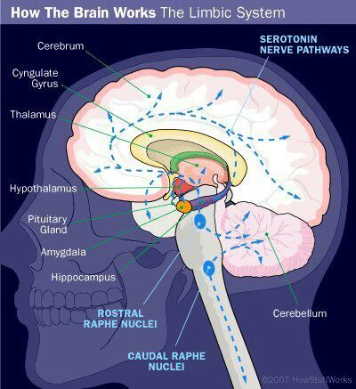 The output of the cortex in these directions during sleep is blocked, and the output signals from the cortex in the state of sleep go to structures associated with associative visceral regulation. It can be assumed that such structures are the nuclei of the hypothalamus. Since people do not have awareness of the visceral sphere, the complexity of the processes of processing visceral information was clearly underestimated, and the central structures of visceral control turned out to be studied much less compared to the structures involved in the analysis of exteroceptive information. No wonder. It can be figuratively imagined that it is as difficult for people to study the organization of the analysis of visceral functions, as it would be difficult for a person with congenital blindness to study the organization of the visual system. Another reason for the lack of knowledge about the organization of the central processing of visceral functions was that most of the physiological research in recent years was carried out on awake animals, when visceral information is not transmitted to the cerebral cortex at all.
 But now, after the discovery of significant symmetry in the organization of the analysis of information flows in wakefulness and in sleep, it has become possible to transfer the patterns discovered in the study of exteroceptive systems to the work of interoceptive systems. The first steps in this direction have already been taken [25].
The visceral theory of sleep also made it possible to approach the analysis of the mechanisms of the transition from wakefulness to sleep. This rather complex system, and its detailed analysis was carried out in the article [25]. Briefly, this process can be represented as a balance between two groups of needs: wakefulness and sleep.
Waking needs are determined by the activity of the organism in the environment and are determined by information coming from the extero- and proprioreceptors. Sleep needs are determined by the state of the visceral systems of the body and the flow of interoceptive information coming from all these organs. If the task of sleep is to restore the body's working capacity, then it is logical to assume that the need for sleep will arise when a mismatch signal (error signal) appears between the genetically determined values of the operation parameters of all visceral systems and the current signals coming from interoreceptors. Until this signal is present, the body remains awake. However, when these parameters begin to deviate from a safe level, this is perceived as drowsiness and the need to go to sleep. We assume that emotional evaluation structures (hippocampus, amygdala, hypothalamus) are involved in this work, and it is these structures, which are really highly active in the sleep state, that send control signals that open or close conduction along different nerve pathways. The places of application of these signals in fig. 1 and 2 are shown with small gray arrows.
The structure of information flows shown in the diagrams will function well under one important condition - all switching will occur strictly synchronously.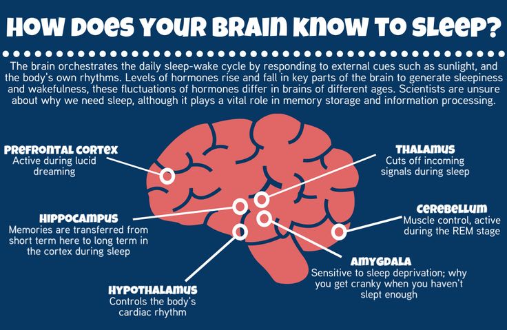 But in reality, these switches are ordinary chemical synapses, and the effectiveness of their work depends on many different influences. Therefore, situations may arise when a particular stream will be opened or closed with a lead or a delay, and as a result, information will go to the wrong address for some time. An analysis of the consequences of such false redirection has shown that many pathological conditions associated with the sleep-wake cycle can be explained by this. Such phenomena as hypnagogic hallucinations, dreams, sleep paralysis, restless legs syndrome, somnambulism are explained by the erroneous casting of visceral information into the blocks of behavior and consciousness. In one of the papers [26], we analyzed each of these cases in detail. Surprisingly, all these cases can be explained by failures in the conduction of signals only in the five switching elements shown in the diagrams. But it must be remembered that in reality, behind each switch shown in the diagram, there are dozens of elements similar in function, distributed over different parts of a living organism, which open up great opportunities for a more detailed analysis of various pathological abnormalities.
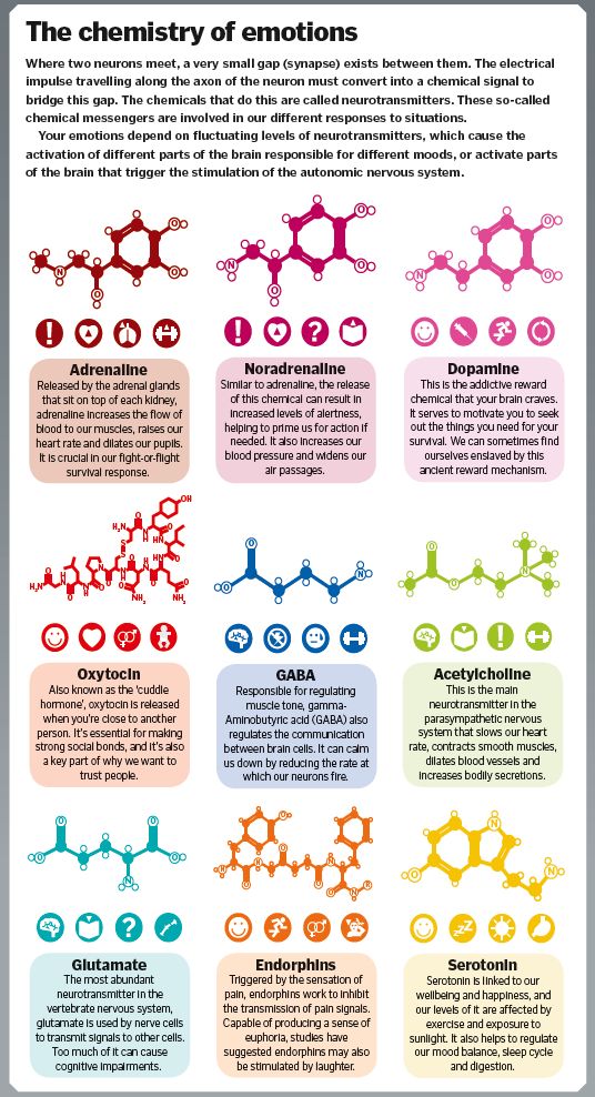
But more frequent and unpleasant consequences can be caused by the throwing of exteroceptive information into the block of associative visceral regulation, leading, for example, to the often encountered phenomenon of motion sickness in transport or visceral problems in people in the first days of entering weightlessness. Most likely, it is failures in the addressing of information flows that underlie psychosomatic diseases. These issues were analyzed in more detail in our previous works [26, 27]. To date, only the very first steps have been taken towards using the visceral theory of sleep to analyze the origins of pathological conditions in the body.
It should be noted that in recent years, the efforts of many somnologists have been directed to studying the consequences of sleep disturbance on the cognitive sphere. Indeed, in many studies, memory deterioration was noted against the background of sleep deprivation, an increase in the number of errors in making complex decisions, and, conversely, an improvement in memorization if learning preceded sleep [28]. These data supported the general belief in the importance of sleep primarily for the functioning of the brain and was sometimes used to criticize the visceral theory of sleep. However, we must remember that the brain is also a visceral organ, and the brain also needs sleep to maintain and restore its functionality. There is nothing surprising in the fact that after sleep the brain begins to work more efficiently. Rather surprisingly, the brain appears to be the most resilient to sleep deprivation. This suggests that there may be special mechanisms in the CNS that constantly maintain a minimum level of brain performance. Its performance may improve during sleep, but when sleep is deprived, its performance does not suffer as much as it happens with other visceral systems.
The provisions of the visceral theory of sleep are not so easy to immediately understand and accept. It is difficult to give up ideas that connect cognitive functions mainly with the cerebral cortex, from cortical fields that have a single and constant function, from permanent connections in the central nervous system, and from the fact that visceral systems can only manage with the control of a small autonomic nervous system.