How to get a brain scan
Brain Scan | Dignity Health
Overview of brain scan
Have you experienced signs and symptoms of a neurological disorder? To confirm a diagnosis, your Dignity Health doctor may order a brain scan. A brain scan can refer to several different types of imaging tests.
Brain scans represent a way for your doctor to “see inside” your skull without having to do surgery. Our neurologists are experts at performing and reading brain scans. To learn more and get started, Find a Doctor today.
Why it’s necessary
As an imaging test, a brain scan can help your doctor evaluate structures within the brain. Some types of tests, like a positron emission tomography (PET) scan, can show how well your brain is functioning. Our neurologists use brain scans to help diagnose or monitor neurological diseases or disorders.
Brain scans are an excellent way to determine neurological diagnoses noninvasively. The information gained from a brain scan will help your doctor treat your neurological condition.
Your doctor will recommend a brain scan if he or she has reason to believe you may have a neurological condition or brain injury, or in order to rule out these conditions.
Common conditions treated with brain scans
Doctors are able to diagnose and monitor a variety of neurological conditions with brain scans, including:
- Brain tumors or cysts
- Blood vessel abnormalities
- Multiple sclerosis
- Stroke
- Epilepsy or hydrocephalus
- Head trauma including concussion>
Talk to your doctor if you have a neurological condition that may need to be monitored with a brain scan
Types
There are three major types of brain scans: computed tomography (CT)magnetic resonance imaging (MRI), and positron emission tomography (PET). Within these categories, there are many other subtypes of brain scans that can measure oxygen usage in the brain, cerebral blood volume, and other aspects of brain structure or function.
CT scan of the brain
This test uses ionizing radiation (X-ray) to produce images of organs, tissues, and bones. CT scans are useful because they can show the area of the brain that is affected and can be used quickly to detect bleeding or clots. The procedure may be performed with or without the use of contrast dye, which is used to highlight different tissues in the brain. In either case, you will be asked to change into a hospital gown and lie on a special table that travels through the narrow opening of the imaging machine. If iodine-containing contrast dye is used, you will receive an intravenous (IV) line. The whole procedure lasts about 20 minutes.
CT scans are used to detect:
- Tumors and cysts
- Brain damage
- Bone or vascular irregularities
- Hydrocephalus (fluid buildup in the brain)
MRI brain scan
Unlike a CT scan, which uses ionizing radiation, MRI uses a magnetic field and radio waves to produce images. You will be asked to change and then lie on a table that moves through the magnetic resonance imaging machine. You cannot have any magnetic metal on or inside your body during an MRI. The most common type of contrast dye for brain MRI is gadolinium — if used, you will receive an IV line. For brain MRIs, a detector is also placed over the head.
MRIs are great for providing:
- Detailed images of tissues
- Blood flow measurements
- Information on any mineral deposits
- Diagnosis of stroke, multiple sclerosis, traumatic brain injury, tumors, infection, inflammation, and more
MRIs are painless and risk-free, though patients who are claustrophobic or obese may be uncomfortable during the procedure. Generally, MRIs take about an hour to complete.
Functional MRIs (fMRI) are used to obtain real-time images of blood flow in the brain. This is especially helpful to pinpoint areas that are active. They can also be used to localize brain areas of motor function, language, or sensation. They are helpful to study degenerative disorders as well.
PET scan of the brain
While CT and MRI scans look at the structure of the brain, a PET scan can show actual brain function. This test involves the injection of a radioactive tracer substance through an IV line in your hand or arm. This material takes about one hour to circulate through your system. After it does, you will lie on a table that slides through the scanning machine. The PET scan uses gamma rays.
PET scans show tumors and diseased tissue, blood flow, and can measure tissue metabolism. They are especially helpful in evaluating patients with epilepsy or memory disorders. PET scans can be used to diagnose cancer, as well as determine how you are responding to your cancer treatment.
PET scans last from 30 minutes to two hours. Some patients find it difficult to lie still for extended periods, while others experience anxiety and discomfort in the enclosed space of the scanning machine. Talk to your doctor or technician if you have claustrophobic tendencies, and about what steps can be taken to make the scan easier for you.
Risks
The risks for brain scans are generally low. CT and PET scans use radiation, but the amount of exposure is quite low and does not last long in your body.
There are heightened risks for pregnant women because fetuses are more sensitive to the effects of radiation. MRIs are a better option for pregnant women. If you are pregnant or think you may be pregnant, you should always discuss this with your doctor before undergoing a diagnostic scan or other procedure.
It is also possible to have an allergic reaction to the radioactive dye. This is very rare but can result in swelling, pain, or redness at the injection site.
The information contained in this article is meant for educational purposes only and should not replace advice from your healthcare provider.
What It Is, Purpose, Procedure & Results
Overview
Healthcare providers order brain MRIs for several different reasons, including to help diagnose new neurological conditions or to monitor existing conditions.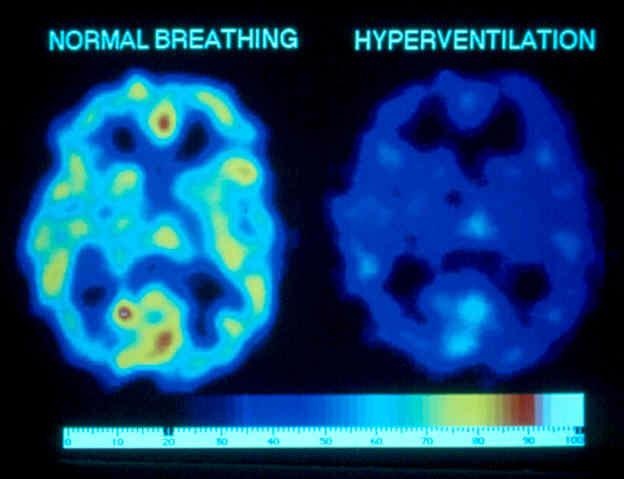
What is a brain MRI?
A brain MRI (magnetic resonance imaging) scan, also called a head MRI, is a painless procedure that produces very clear images of the structures inside of your head — mainly, your brain. MRI uses a large magnet, radio waves and a computer to produce these detailed images. It doesn’t use radiation.
Currently, MRI is the most sensitive imaging test of your head (particularly, your brain), as compared to other imaging techniques, such as CT (computed tomography) scans or X-rays.
What is a brain MRI with contrast?
Some brain MRI exams use an injection of contrast material. The contrast agent is often gadolinium, which is a rare earth metal. When this substance is present in your body, it alters the magnetic properties of nearby water molecules, which enhances the quality of the images. This improves the sensitivity and specificity of the diagnostic images.
Contrast material enhances the visibility of the following:
- Tumors.

- Inflammation.
- Certain organs’ blood supply.
- Blood vessels.
The contrast can also help diagnose multiple sclerosis, stroke, dementia and infection.
If your brain MRI requires a contrast material, your healthcare provider will insert an intravenous catheter (IV line) into a vein in your hand or arm. They’ll use this IV to inject the contrast material.
Contrast materials are safe intravenous (IV) drugs. Side effects, ranging from mild to severe, do occur, but severe reactions are very rare.
What is the difference between a head MRI and a brain MRI?
A head MRI and a brain MRI are the same procedure. They both provide images of the inside of your head. While healthcare providers most often use head and brain MRIs to assess your brain, these imaging procedures provide images of other structures in your head, too, such as facial bones, blood vessels and nerves.
What does a brain MRI show?
A brain or head MRI shows the structures inside of your head, including:
- Your brain.
- Blood vessels that connect to your brain.
- Your skull and facial bones.
- Structures in your inner ear.
- Your eyes and their supporting tissues, such as your optic nerves.
- Other nerves (large nerves in your head, called cranial nerves).
- Surrounding soft tissues and skull-based structures, such as fat, bones, muscle and connective tissue.
More specifically, a brain or head MRI can show if there are any abnormalities in your brain or the surrounding tissues, including, but not limited to:
- Inflammation and swelling.
- Structural issues.
- Abnormal growths or masses.
- Fluid leaks.
- Hemorrhage (bleeding inside your brain).
- White matter disease.
Why would a neurologist order an MRI of the brain?
Neurologists and other healthcare providers order brain MRIs for several different reasons, including helping diagnose new neurological conditions based on certain symptoms or to monitor existing conditions.
Some of the conditions a brain MRI can help diagnose or monitor include:
- A blood clot in your brain.
- Brain aneurysm.
- Brain hemorrhage.
- Brain infections (encephalitis).
- Brain damage associated with epilepsy.
- Brain tumors and cysts.
- Certain chronic neurological conditions, such as multiple sclerosis (MS).
- Dementia.
- Hydrocephalus.
- Pituitary gland issues, such as a pituitary adenoma.
- Stroke.
- Issues with brain development or structure, such as Chiari malformation, and malformations of cortical development. (The term “cortical” refers to the outer layer of your cerebrum.)
- Traumatic brain injury (TBI).
Your healthcare provider may also order an MRI of your head if you have any combination of the following signs and symptoms:
- Migraines and/or chronic headaches.
- Seizures.
- Vertigo and frequent episodes of severe dizziness.
- Hearing loss with an unexplainable cause.
- Vision issues not explained by an eye exam.
- Hormonal imbalances related to your hypothalamus and/or pituitary gland.
- Significant changes to your thinking and behavior
- Extreme weakness and fatigue.
Healthcare providers also use brain and head MRI scans before surgeries involving your head to better prepare for the surgery. They also use these scans to ensure that healing from the surgery is going well. Any significant injuries involving your head also prompt healthcare providers to order brain MRI scans to check for injuries, bleeding and swelling.
Who performs a brain MRI?
A radiologist or a radiology technologist will perform your brain (head) MRI. A radiologist is a medical doctor who performs and interprets imaging tests to diagnose conditions. A radiology technologist is a healthcare provider who’s specially trained and certified to perform an MRI scan.
Test Details
How does a brain MRI work?
Magnetic resonance imaging (MRI) works by passing an electric current through coiled wires to create a temporary magnetic field in your body — in this case, your head.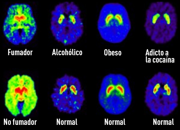 A transmitter/receiver in the machine then sends and receives radio waves. The computer then uses these signals to make digital images of the structures inside of your head, including your brain.
How do I prepare for a brain MRI?
Guidelines about eating and drinking before a brain MRI vary based on the reason for your MRI. Eat and take your medications as usual unless your healthcare provider tells you otherwise.
The magnetic resonance imaging (MRI) scanner uses strong magnets and radio wave signals that can cause heating or possible movement of some metal objects in your head and/or body. This could result in health and safety issues. It could also cause some implanted electronic medical devices to malfunction.
If you have metal-containing objects or implanted medical devices in your body, your healthcare provider needs to know about them before your brain MRI. Certain implanted objects may require additional scheduling arrangements and special instructions. Other items don’t require special instructions but may require an X-ray to check on the exact location of the object before your exam.
It’s important to tell your healthcare provider and MRI technologist if you have any of the following:
- Cardiac pacemaker.
- Middle ear prostheses.
- Cochlear implant.
- A clip used for brain aneurysms.
- Vagal nerve stimulator.
- Metal fragments in your head or within your eyeball.
In addition, tell your healthcare provider if you:
- Are pregnant.
- Aren’t able to lie on your back for 30 to 60 minutes.
- Have claustrophobia (fear of enclosed or narrow spaces).
Leave all jewelry and other accessories at home or remove them before your brain MRI. Metal and electronic items aren’t allowed in the exam room because they can interfere with the magnetic field of the MRI unit, cause burns or become harmful projectiles. These items include:
- Jewelry, watches, credit cards and hearing aids — all of which can be damaged.
- Pins, metal hair accessories, underwire bras and metal zippers, which can distort MRI images.
- Removable dental work, such as dentures.
- Pens, pocketknives and eyeglasses.
- Body piercings.
- Cell phones, electronic watches and tracking devices.
What should I expect during a brain MRI?
Most brain MRI exams are painless, but some people find it uncomfortable to remain still for 30 minutes or longer. Others may experience anxiety due to the closed-in space while in the MRI machine. The machine can also be noisy.
The general steps of a brain MRI scan and what to expect include:
- You’ll change into a hospital gown for the MRI scan.
- You’ll lie face up for most exams on the MRI scanning bed.
- Once you’re lying on the table, the technologist will position a special helmet-like device called a head coil around your head. Some head coils have a mirror attached to them that allows you to see outside of the scanning machine or a small screen that allows you to watch television. This can help prevent feelings of claustrophobia.
- The technologist will then slide you and the scanning bed into the MRI machine.
- As the MRI scan begins, you’ll hear the equipment making a variety of loud knocking and clicking sounds while it’s taking the images. Each series of sounds may last for several minutes. You’ll be given earplugs or headphones to wear to protect your hearing before the procedure begins. You may also be able to listen to music through the headphones.
- It’s important to be very still during the exam to ensure the best quality of images.
- It’s normal for the area of your body being imaged to feel slightly warm. If it bothers you, tell the radiologist or technologist.
- The MRI technologist will be able to see you and can talk with you at all times. An intercom system allows two-way communication while you’re inside the scanner. You’ll also have a call button in your hand that you can push to let the technologist know if you’re having any problems or concerns.
In some cases, your MRI may require contrast. If this applies to you, your healthcare provider will give you an IV injection of contrast material before you undergo the MRI. The IV needle may cause some discomfort but this won’t last long. You may have some bruising afterward. Some people experience a temporary metallic taste in their mouth after the contrast injection.
If you have claustrophobia, your healthcare provider may recommend a sedative drug so you feel more relaxed during the exam, or even anesthesia.
Does your whole body go into the machine for a brain MRI?
In most cases, your whole body won’t go into the MRI machine tunnel if you’re only getting a head or brain MRI.
How long does a brain MRI take?
A brain MRI can take about 30 minutes to an hour to complete. It may take longer if you’re getting a brain MRI with contrast.
Your healthcare provider will be able to give you a more exact time range based on the specific reason for your scan.
Results and Follow-Up
When should I know the results of the test?
After your MRI scan, a radiologist will analyze the images. The radiologist will send a signed report to your primary healthcare provider, who will share the results with you. The report is usually ready for your healthcare provider within one or two days.
You may need a follow-up exam. If so, your healthcare provider will explain why.
A note from Cleveland Clinic
Brain magnetic resonance imaging (MRI) is a very useful and generally safe imaging test that healthcare providers use for a variety of reasons. If you need a brain MRI scan and are worried about the exam or have questions about it, don’t hesitate to ask your healthcare provider. They’re available to answer your questions and support you.
Dopplerography and duplex scanning of blood vessels — FSBI "NMITs TPM" of the Ministry of Health of Russia
Dopplerography and duplex scanning - two related methods of ultrasound examination of blood vessels.
The essence and differences of the methods
Dopplerography of the vessels of the brain, neck, upper and lower extremities, as well as their duplex scanning, belongs to non-invasive diagnostic procedures. Their advantage is affordable cost and the absence of contraindications, high information content.
Using the Doppler effect allows you to calculate the speed of blood flow, to determine its violation in individual vessels. Most often, these data are enough for the doctor to make an accurate diagnosis. In turn, duplex scanning of the vessels of the neck, head and extremities provides information not only on the quality of blood flow, but also on the geometry of the vascular lumen, the tortuosity of the channel, the presence of anatomical or postoperative anomalies, wall thickness, the appearance of blood clots and atherosclerotic plaques.
GNITsPM offers to use the possibilities of modern ultrasound diagnostics as part of a comprehensive or routine examination.
Indications for ultrasound diagnostics of blood vessels
Dopplerography of the vessels of the brain and other organs is appropriate as a diagnostic tool for routine preventive examinations, when the likelihood of serious problems is low. In patients with osteochondrosis, dopplerography of the vessels of the head and neck makes it possible to identify the effect of the disease on the circulatory system. Timely Dopplerography of the vessels of the lower extremities is important in making such diagnoses as:
- varicose disease;
- atherosclerosis obliterans and endarteritis;
- deep vein thrombosis.
Since duplex scanning is more informative, it is effective for clarifying the diagnosis. Duplex scanning of cerebral vessels is prescribed in the same cases as conventional dopplerography, as well as if it is necessary to localize the problem area. This study is recommended for everyone over the age of 40, and visitors to our medical center are increasingly taking advantage of this opportunity.
Scanning of the brachiocephalic arteries plays an important role in the prevention of such a dangerous disorder as a stroke. It is prescribed for:
- headaches or dizziness of unknown origin;
- planning of surgical intervention in the cardiovascular system;
- examination of people at risk of cerebrovascular accident;
- the presence of symptoms of developing stroke or compression of the area of the brachiocephalic arteries.
Duplex scanning of the arteries of the lower extremities, as well as scanning of the veins , , gives the specialist a detailed picture of the state of the vessels. It not only displays the presence of blood flow disorders, but also explains their cause, whether it be vascular anomalies, the consequences of injuries, atherosclerotic changes, or something else. Most often, they turn to us in the direction of a phlebologist in order to scan the veins of the lower extremities.
If you have ever noticed:
- swollen legs in the evening;
- discoloration of the skin, appearance of veins;
- numbness and muscle pain when walking;
- cold feet, -
then a scan of the vessels of the lower extremities will clarify the nature of your condition, which means it will help you start corrective therapy in time.
This page is for informational purposes only. The exact list of services provided and the features of the procedures can be found by phone.
Where to do and what the duplex scan shows
- Main
- Blog
- Where to do a duplex scan
Content:
-
What a duplex vascular scan shows
-
How duplex scanning is performed
-
How duplex scanning of the head, peritoneum and legs works
-
Which is better: duplex scanning or MRI
-
Where can I get a duplex scan
Ultrasonography plays a key role in the diagnosis of vascular diseases. To obtain a more accurate picture, they resort to the method of double duplex scanning (USDS). What does duplex scanning mean? The method combines two types of scanning - ultrasound of adjacent tissues and vascular Doppler. From the article, you can learn how duplex scanning of the vessels of the head is done, how duplex scanning of the abdominal vessels is performed, and how duplex scanning of the lower extremities is done.
What a duplex vascular scan shows
This technique is safe, non-invasive and painless. Ultrasound can be performed on both infants and pregnant women.
What duplex scanning shows:
- condition of the arteries and vessels of the spine, abdomen, lower extremities, main veins responsible for nourishing the brain, neck, upper extremities;
- the appearance of plaques, blood clots, the degree of narrowing of the lumen of the vessel, the speed of blood flow, the patency of veins and arteries, the expansion of the vessel;
- condition of veins and vessels after operations and injuries;
- congenital developmental defects.
Indications for the appointment of the procedure are various conditions: recurring dizziness, fainting, varicose veins, suspected atherosclerosis, aneurysm, control before surgery.
How duplex scanning is performed
Examination time does not take more than 30-50 minutes. Usually no preparation is required when examining the head or extremities. Preparation is required only when examining the abdominal cavity.
How a duplex scan is done: The patient is asked to expose the area to be examined and assume a supine or sitting position. Sometimes an ultrasound specialist may ask you to strain one or another organ.
How does duplex scanning of the head, peritoneum and legs work? , then the procedure as a whole does not differ from a conventional ultrasound.
What a duplex scan of the lower extremities shows: during ultrasound, you can detect thrombosis of the saphenous and deep veins, determine the cause of swelling, monitor the condition of the vessels before surgery. veins, sometimes the patient may be asked to roll over on his stomach, sit down or stand up. Below we will tell you where to do a duplex scan of the veins of the lower extremities.
How is duplex scanning of the abdominal cavity performed: three days before the examination, you need to follow a diet, do not eat foods that cause gas formation. Gas-reducing drugs and cleansing enemas may be prescribed. Do not eat, drink caffeinated drinks or smoke before the procedure.
Which is better: duplex scanning or MRI
You have learned how duplex vein scanning is done, but you are probably wondering whether to choose ultrasound or MRI. It is impossible to say unequivocally which research method is better. Both methods are widely used and successful. Ultrasound diagnostics has no contraindications. No special preparation is required before the examination. Dopplerography is quite informative, moreover, it is a more accessible procedure than MRI. Many places have now opened where duplex scanning of blood vessels can be done, but it is better to contact an advanced center where the diagnosis will be carried out by a practicing doctor, for example, the Chekhov Vascular Center.
MRI is more often prescribed for already diagnosed pathologies. Tomography helps to obtain a three-dimensional detailed image of the vessels and establish not only the presence of a problem, but also the cause and degree of the disease. MRI is not prescribed in the presence of metal structures in the body, as well as in case of claustrophobia. You need to be completely immobilized during a tomography, so this method is not suitable for diagnosing infants. Also, MRI is not prescribed during pregnancy. In this regard, ultrasound is a more gentle technique with zero radiation exposure.
Where to get a duplex scan
If you are looking for where to get a duplex scan, go to a clinic that has an ultrasound room. The quality of data interpretation also depends on the qualifications of the diagnostician. If you are interested in where you can get a duplex vein scan with the most accurate result, contact the Chekhov Vascular Center - we have surgeons, ultrasound diagnostics specialists who are knowledgeable about the latest advances in medical technology.
Now you know how duplex scanning of vessels is done and where to do duplex scanning of veins. If you notice the first symptoms of cardiovascular disease in yourself, do not delay visiting a doctor.