What techniques are used to view or measure the frontal lobe
A systematic review of brain frontal lobe parcellation techniques in magnetic resonance imaging
Review
. 2014 Jan;219(1):1-22.
doi: 10.1007/s00429-013-0527-5. Epub 2013 Mar 10.
Simon R Cox 1 , Karen J Ferguson, Natalie A Royle, Susan D Shenkin, Sarah E MacPherson, Alasdair M J MacLullich, Ian J Deary, Joanna M Wardlaw
Affiliations
Affiliation
- 1 Brain Research Imaging Centre, University of Edinburgh, Edinburgh, UK, [email protected].
- PMID: 23474540
- DOI: 10.
1007/s00429-013-0527-5
Free article
Review
Simon R Cox et al. Brain Struct Funct. 2014 Jan.
Free article
. 2014 Jan;219(1):1-22.
doi: 10.1007/s00429-013-0527-5. Epub 2013 Mar 10.
Authors
Simon R Cox 1 , Karen J Ferguson, Natalie A Royle, Susan D Shenkin, Sarah E MacPherson, Alasdair M J MacLullich, Ian J Deary, Joanna M Wardlaw
Affiliation
- 1 Brain Research Imaging Centre, University of Edinburgh, Edinburgh, UK, simon.
 [email protected].
- PMID: 23474540
- DOI: 10.1007/s00429-013-0527-5
Abstract
Manual volumetric measurement of the brain's frontal lobe and its subregions from magnetic resonance images (MRIs) is an established method for researching neural correlates of clinical disorders or cognitive functions. However, there is no consensus between methods used to identify relevant boundaries of a given region of interest (ROI) on MRIs, and those used may bear little relation to each other or the underlying structural, functional and connective architecture. This presents challenges for the analysis and synthesis of such results. We therefore performed a systematic literature review to highlight variations in the anatomical boundaries used to measure frontal regions, contextualised by up-to-date evidence from histology, hodology and neuropsychology. We searched EMBASE and MEDLINE for studies in English reporting three-dimensional boundaries for manually delineating the brain's frontal lobe or sub-regional ROIs from MRIs. Exclusion criteria were: exclusive use of co-ordinate grid systems; insufficient detail to allow method replication; publication in grey literature only. Papers were assessed on quality criteria relating to bias, reproducibility and protocol rationale. There was a large degree of variability in the three-dimensional boundaries of all regions used by the 208 eligible papers. Half of the reports did not justify their rationale for boundary selection, and each paper met on average only three quarters of quality criteria. For the frontal lobe and each subregion (frontal pole, anterior cingulate, dorsolateral, inferior-lateral, and orbitofrontal) we identified reproducible methods for a biologically plausible target ROI. It is hoped that this synthesis will guide the design of future volumetric studies of cerebral structure.
Similar articles
-
Automated MRI parcellation of the frontal lobe.
Ranta ME, Chen M, Crocetti D, Prince JL, Subramaniam K, Fischl B, Kaufmann WE, Mostofsky SH. Ranta ME, et al. Hum Brain Mapp. 2014 May;35(5):2009-26. doi: 10.1002/hbm.22309. Epub 2013 Jul 29. Hum Brain Mapp. 2014. PMID: 23897577 Free PMC article.
-
Human frontal cortex: an MRI-based parcellation method.
Crespo-Facorro B, Kim JJ, Andreasen NC, O'Leary DS, Wiser AK, Bailey JM, Harris G, Magnotta VA. Crespo-Facorro B, et al. Neuroimage. 1999 Nov;10(5):500-19. doi: 10.1006/nimg.1999.0489. Neuroimage. 1999. PMID: 10547328
-
Manual MRI parcellation of the frontal lobe.
Ranta ME, Crocetti D, Clauss JA, Kraut MA, Mostofsky SH, Kaufmann WE. Ranta ME, et al. Psychiatry Res. 2009 May 15;172(2):147-54. doi: 10.1016/j.pscychresns.2009.01.006. Epub 2009 Mar 25. Psychiatry Res. 2009. PMID: 19324532 Free PMC article.
-
Architecture and organizational principles of Broca's region.
Amunts K, Zilles K. Amunts K, et al. Trends Cogn Sci. 2012 Aug;16(8):418-26. doi: 10.1016/j.tics.2012.06.005. Epub 2012 Jul 3. Trends Cogn Sci. 2012. PMID: 22763211 Review.
-
Large-Scale Meta-Analysis of Human Medial Frontal Cortex Reveals Tripartite Functional Organization.
de la Vega A, Chang LJ, Banich MT, Wager TD, Yarkoni T. de la Vega A, et al. J Neurosci. 2016 Jun 15;36(24):6553-62.
 doi: 10.1523/JNEUROSCI.4402-15.2016. J Neurosci. 2016. PMID: 27307242 Free PMC article. Review.
See all similar articles
Cited by
-
Radiation dose to circumscribed brain regions and neurocognitive function in patients with meningioma.
Sekely A, Tsang DS, Mabbott D, Kongkham P, Zadeh G, Zakzanis KK, Edelstein K. Sekely A, et al. Neurooncol Pract. 2022 Feb 19;9(3):208-218. doi: 10.1093/nop/npac011. eCollection 2022 May. Neurooncol Pract. 2022. PMID: 35601975
-
Increased Homotopic Connectivity in the Prefrontal Cortex Modulated by Olanzapine Predicts Therapeutic Efficacy in Patients with Schizophrenia.
Shan X, Liao R, Ou Y, Ding Y, Liu F, Chen J, Zhao J, He Y, Guo W.
 Shan X, et al. Neural Plast. 2021 Sep 1;2021:9954547. doi: 10.1155/2021/9954547. eCollection 2021. Neural Plast. 2021. PMID: 34512748 Free PMC article. Clinical Trial.
 -
Reduced Temporal Activation During a Verbal Fluency Task is Associated with Poor Motor Speed in Patients with Major Depressive Disorder.
Kiriyama T, Tanemura R, Nakamura Y, Takemoto C, Hashimoto M, Utsumi H. Kiriyama T, et al. Psychiatry Investig. 2020 Aug;17(8):804-813. doi: 10.30773/pi.2020.0045. Epub 2020 Aug 18. Psychiatry Investig. 2020. PMID: 32791821 Free PMC article.
-
Psychotic-like experiences, polygenic risk scores for schizophrenia, and structural properties of the salience, default mode, and central-executive networks in healthy participants from UK Biobank.
Alloza C, Blesa-Cábez M, Bastin ME, Madole JW, Buchanan CR, Janssen J, Gibson J, Deary IJ, Tucker-Drob EM, Whalley HC, Arango C, McIntosh AM, Cox SR, Lawrie SM. Alloza C, et al. Transl Psychiatry. 2020 Apr 27;10(1):122. doi: 10.1038/s41398-020-0794-x. Transl Psychiatry. 2020. PMID: 32341335 Free PMC article.
-
Fluctuating asymmetry in brain structure and general intelligence in 73-year-olds.
Moodie JE, Ritchie SJ, Cox SR, Harris MA, Muñoz Maniega S, Valdés Hernández MC, Pattie A, Corley J, Bastin ME, Starr JM, Wardlaw JM, Deary IJ. Moodie JE, et al. Intelligence. 2020 Jan-Feb;78:101407. doi: 10.1016/j.intell.2019.101407. Intelligence. 2020. PMID: 31983789 Free PMC article.
See all "Cited by" articles
Publication types
MeSH terms
Grant support
- BB_/Biotechnology and Biological Sciences Research Council/United Kingdom
- G0700704/MRC_/Medical Research Council/United Kingdom
- G1001245/MRC_/Medical Research Council/United Kingdom
- G0700704/84698/MRC_/Medical Research Council/United Kingdom
- MR/K026992/1/MRC_/Medical Research Council/United Kingdom
Measurement of frontal lobe volume on magnetic resonance imaging scans
. 1997 Aug 8;75(1):23-30.
doi: 10.1016/s0925-4927(97)00026-7.
E H Aylward 1 , A Augustine, Q Li, P E Barta, G D Pearlson
Affiliations
Affiliation
- 1 Department of Psychiatry and Behavioral Sciences, Johns Hopkins University School of Medicine, Baltimore, MD 21287-7362, USA.
- PMID: 9287371
- DOI: 10.1016/s0925-4927(97)00026-7
E H Aylward et al. Psychiatry Res. .
. 1997 Aug 8;75(1):23-30.
doi: 10.1016/s0925-4927(97)00026-7.
Authors
E H Aylward 1 , A Augustine, Q Li, P E Barta, G D Pearlson
Affiliation
- 1 Department of Psychiatry and Behavioral Sciences, Johns Hopkins University School of Medicine, Baltimore, MD 21287-7362, USA.
- PMID: 9287371
- DOI: 10.1016/s0925-4927(97)00026-7
Abstract
This article describes rules for measurement of the frontal lobe on thin SPGR (spoiled gradient recalled echo in steady state) MRI (magnetic resonance imaging) scans. Measurements were performed using a locally-developed software program that allows 3-dimensional reconstruction of images, 'painting' of landmarks on the surface of the brain, and reconstruction of 2-dimensional images in any plane with landmark 'paint' remaining on the surface of the brain. Excellent inter-rater reliability has been achieved for this method. The approach may be particularly useful for studies involving groups of patients whose brains are known to be dysmorphic and who may not, therefore, be appropriate for measurement methods that involve image warping or dependence on arbitrary landmarks for defining the posterior boundary of the frontal lobe.
Similar articles
-
[Quantitative measurement of prefrontal lobe volume on three dimensional magnetic resonance imaging scan].
Kanemura H, Aihara M, Aoki S, Hatakeyama K, Kamiya Y, Ono C, Sata Y, Nakazawa S.
 Kanemura H, et al. No To Hattatsu. 1999 Nov;31(6):519-24. No To Hattatsu. 1999. PMID: 10565188 Japanese.
 -
Reliability and validity of MRI measurement of the amygdala and hippocampus in children with fragile X syndrome.
Kates WR, Abrams MT, Kaufmann WE, Breiter SN, Reiss AL. Kates WR, et al. Psychiatry Res. 1997 Aug 8;75(1):31-48. doi: 10.1016/s0925-4927(97)00019-x. Psychiatry Res. 1997. PMID: 9287372
-
Landmark-based morphometrics of the normal adult brain using MRI.
Free SL, O'Higgins P, Maudgil DD, Dryden IL, Lemieux L, Fish DR, Shorvon SD. Free SL, et al. Neuroimage. 2001 May;13(5):801-13. doi: 10.1006/nimg.2001.0748. Neuroimage. 2001. PMID: 11304077
-
Automatic atlas-based volume estimation of human brain regions from MR images.
Andreasen NC, Rajarethinam R, Cizadlo T, Arndt S, Swayze VW 2nd, Flashman LA, O'Leary DS, Ehrhardt JC, Yuh WT. Andreasen NC, et al. J Comput Assist Tomogr. 1996 Jan-Feb;20(1):98-106. doi: 10.1097/00004728-199601000-00018. J Comput Assist Tomogr. 1996. PMID: 8576490
-
A systematic review of brain frontal lobe parcellation techniques in magnetic resonance imaging.
Cox SR, Ferguson KJ, Royle NA, Shenkin SD, MacPherson SE, MacLullich AM, Deary IJ, Wardlaw JM. Cox SR, et al. Brain Struct Funct. 2014 Jan;219(1):1-22. doi: 10.1007/s00429-013-0527-5. Epub 2013 Mar 10. Brain Struct Funct. 2014. PMID: 23474540 Review.
See all similar articles
Cited by
-
Patterns of differences in brain morphology in humans as compared to extant apes.
Aldridge K. Aldridge K. J Hum Evol. 2011 Jan;60(1):94-105. doi: 10.1016/j.jhevol.2010.09.007. Epub 2010 Nov 5. J Hum Evol. 2011. PMID: 21056456 Free PMC article.
-
Magnetic resonance imaging outcomes from a comprehensive magnetic resonance study of children with fetal alcohol spectrum disorders.
Astley SJ, Aylward EH, Olson HC, Kerns K, Brooks A, Coggins TE, Davies J, Dorn S, Gendler B, Jirikowic T, Kraegel P, Maravilla K, Richards T. Astley SJ, et al. Alcohol Clin Exp Res. 2009 Oct;33(10):1671-89. doi: 10.1111/j.1530-0277.2009.01004.x. Epub 2009 Jul 1. Alcohol Clin Exp Res. 2009. PMID: 19572986 Free PMC article.
-
Manual MRI parcellation of the frontal lobe.
Ranta ME, Crocetti D, Clauss JA, Kraut MA, Mostofsky SH, Kaufmann WE.
 Ranta ME, et al. Psychiatry Res. 2009 May 15;172(2):147-54. doi: 10.1016/j.pscychresns.2009.01.006. Epub 2009 Mar 25. Psychiatry Res. 2009. PMID: 19324532 Free PMC article.
 -
Volumetric neuroimage analysis extensions for the MIPAV software package.
Bazin PL, Cuzzocreo JL, Yassa MA, Gandler W, McAuliffe MJ, Bassett SS, Pham DL. Bazin PL, et al. J Neurosci Methods. 2007 Sep 15;165(1):111-21. doi: 10.1016/j.jneumeth.2007.05.024. Epub 2007 May 29. J Neurosci Methods. 2007. PMID: 17604116 Free PMC article.
-
Relationship of brain and skull in pre- and postoperative sagittal synostosis.
Aldridge K, Kane AA, Marsh JL, Yan P, Govier D, Richtsmeier JT. Aldridge K, et al. J Anat. 2005 Apr;206(4):373-85.
 doi: 10.1111/j.1469-7580.2005.00397.x. J Anat. 2005. PMID: 15817105 Free PMC article.
See all "Cited by" articles
Publication types
MeSH terms
Grant support
- NINDS 16375/DS/DS NIH HHS/United States
- RR00722/RR/NCRR NIH HHS/United States
Test battery for frontal dysfunction in the diagnosis of dementia go to the Memini website
Dementia can be associated with damage to various areas of the brain. In this case, the involvement of a particular site will affect the nature of the symptoms. Therefore, experts create different methods, each of which is more sensitive to a particular disease or its stage. As the name suggests, the frontal dysfunction battery is used to screen for dementias that predominantly affect the frontal lobes or subcortical cerebral structures. In these situations, the sensitivity of the most common cognitive assessment test, the MMSE, may be insufficient.
1. Categorization (generalization).
Question to the patient: What do bananas and oranges have in common?
If the patient finds it difficult ( Nothing in common , Both have peel ...), they suggest the correct answer ( This is fruit ), but the point is not awarded.
Then they ask:
What do a table and a chair have in common?
What do tulips, roses and lilies of the valley have in common?
Each categorical generalization (fruits, furniture, flowers) is worth 1 point. Any other answer is considered incorrect. The correct answer is given only in the first question.
Score: Maximum score - 3, minimum - 0.
2. Speech activity (flexibility of thinking).
Instruction to the patient: Within one minute, name as many words as you can beginning with the letter C . Proper names do not count.
If the patient does not respond in the first 5 seconds, he receives a prompt: For example, the dog is .
If the silence lasts longer than 10 seconds, the doctor repeats the instruction: Any word that begins with the letter "S".
Score: More than 9 words per minute - 3 points, 6 to 9 words - 2 points, 3 to 5 words - 1 point, less than 3 words - 0 points.
3. Dynamic praxis.
Instruction to the patient: Look carefully what I will do.
After that, the doctor, sitting opposite the patient, performs a series of three movements with his left hand three times: fist (placed horizontally, parallel to the table surface) - rib (hand placed vertically on the medial edge) - palm (hands are placed horizontally with the palm up).
After the demonstration of the first three series, the patient is instructed: Now repeat with me with your right hand.
Doctor and patient perform three series together. The instruction follows: Now do the series yourself.
Both during memorization of movements and in the future, verbal prompts are unacceptable (for example, you cannot give a verbal instruction “fist-rib-palm” or say “so-so-and so”).
Evaluation: The patient correctly completed six series in a row on his own - 3 points, correctly completed three series in a row on his own - 2 points, the patient cannot perform the series on his own, but performs them correctly together with the doctor - 1 point, the patient is unable to perform three consecutive series series even with a doctor - 0 points.
4. Simple selection reaction.
The patient is instructed: We will tap the rhythm. If I hit once, you must hit twice in a row.
To make sure the patient understands the instructions correctly, the doctor performs a series of three single blows
If I hit twice in a row, you must hit only once .
To make sure the patient understands the instructions correctly, the doctor performs a series of three double strokes.
Then the following rhythm is tapped: 1-1-2-1-2-2-2-1-1-2.
Evaluation of the result: correct execution - 3 points, no more than 2 errors - 2 points, more than 2 errors - 1 point, complete copying of the doctor's rhythm 4 times in a row or more - 0 points.
5. Complicated choice reaction.
Instruction to the patient: Now if I hit once, you must hit only once.
To make sure the patient understands the instructions correctly, the doctor performs a series of three single blows
If I hit twice in a row, you don't have to do anything
To make sure the patient understands the instructions correctly, the doctor performs a series of three double strokes.
Then the same rhythm is tapped: 1-1-2-1-2-2-2-1-1-2.
The evaluation of the result is similar to item 4.
6. Study of grasping reflexes.
The doctor sits in front of the patient. The patient's hands are on their knees, palms up. Without saying anything or looking at the patient, the doctor brings his hands up and touches both palms.
Evaluation: Lack of grasping reflex - 3 points. The patient hesitates and asks what he should do - 2 points. The patient grabs the doctor's hand - he is instructed not to do this and the grasping reflex is checked again. If the reflex is absent during the re-examination, 1 point is given, otherwise - 0 points.
Interpretation of the result
The threshold interval for distinguishing frontal and Alzheimer's dementias is 12 points. A score below 12 is highly likely to indicate frontal dementia.
Sources:
Dubois B., Litvan I. The FAB: A frontal assessment battery at bedside. Neurology 2000; 55(11): 1621-1626.
Slachevsky A., Dubois B. Frontal assessment battery and differential diagnosis of frontotemporal dementia and Alzheimer disease. Archives of Neurology 2004; 61(7): 1104-1107.
Frontal Dysfunction Test Battery.pdf
Download leaflet
Warning
If you experience similar symptoms, consult your doctor. Do not self-medicate - it is dangerous for your health!how a woman's brain differs from a man's and why a photo of an ex activates areas in the brain associated with physical pain
An fMRI can highlight certain areas of the brain that are activated when people look at pictures of their loved ones. The brains of men and women are differently active before and after sex. A tumor in a certain area of the brain can completely change a person's sexual behavior and turn a school teacher into a maniac. Knowing all this, will it be possible to manipulate falling in love in the future? Polina Krivykh, a clinical psychologist, psychophysiologist, popularizer of science, lecturer and mentor of the School of Lecturers of the Evolution Foundation, spoke about all this.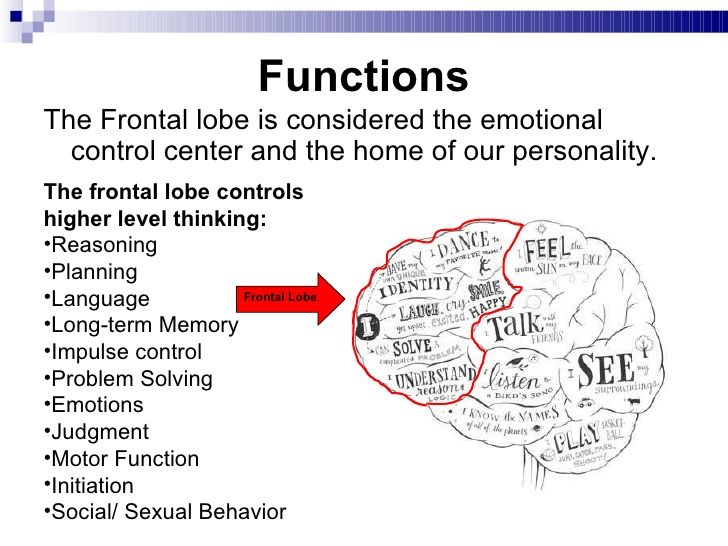
Neurotransmitters and hormones
The variety of “Love is…” pictures suggests that we know nothing about love. The only closest thing to science would be this wording on the gum insert: love is a dopaminergic goal-setting motivation to form pair bonds. Dopamine is a neurotransmitter that is used to communicate between neurons in the brain. How is dopamine related to love? This is easy to understand with the example of monogamous mice-voles, which are absolutely faithful to their partner.
Voles have two types of dopamine receptors in the nucleus accumbens. Thus, D2 type receptors are responsible for the formation of tender attachment to another vole mouse. When a male monogamous vole meets a female and falls in love, his dopamine D2 receptors are activated and an affectionate attachment to a particular female is formed. If these receptors are blocked, the male will bite the females, and if sharply activated, he will fall in love with the first one that comes across.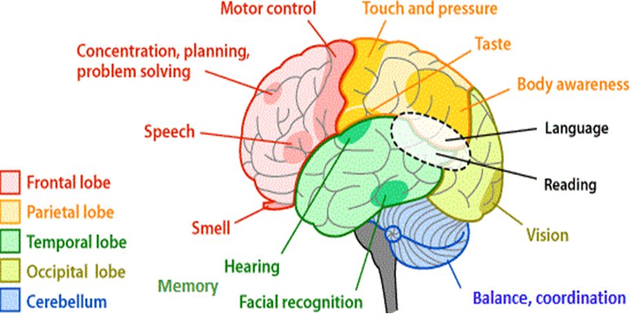 After attachment has formed in the male brain, an increase in the number of D1 receptors occurs, this process reinforces love due to a real “restructuring” of the brain (there are more receptors). The conclusion is simple: after falling in love, the brain of a male vole is rewired so that he can no longer love anyone else. The nucleus accumbens is the same region in which, due to a different way of activating dopamine receptors, addiction to certain drugs arises, so love is often compared to a drug trance. Non-monogamous voles initially have more D1 receptors, meaning they don't need to grow new ones, they have enough of them, so they can switch from one female to another.
There is a serotonin-dopamine balance in the human brain: the higher the serotonin, the less dopamine and vice versa. If a person takes antidepressants (and most antidepressants work by increasing serotonin levels), then dopamine levels decrease, and the person experiences less attraction to the object of their affection, even if the couple is stable.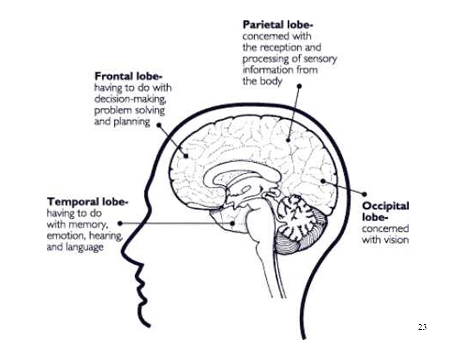
If we transfer the situation to a person, then we should look not at dopamine, but at the hormones vasopressin and oxytocin - substances that are produced in the brain, but then enter the main bloodstream and spread throughout the body. Using the example of monogamous voles, scientists have learned that men have a fidelity gene (women also have it, but it manifests itself differently), it is called RS3334. The same gene is responsible for the formation of vasopressin V1a receptors. This gene can turn monogamous voles into polygamous ones. It is not yet fully understood how this happens, but if the gene is activated and the receptors mutate (that is, the formation of additional V1a receptors), then even a monogamous vole with a large number of such receptors becomes polygamous.
In humans, the situation is similar. A number of interesting studies have shown that men who express such a gene with a particular mutation are unfaithful. However, genetic tests have not yet become widespread, so the results and the name of the gene are based on a couple of Swedish studies, the sample of which is not very large (about 50 people). Believe it or not, the choice is yours.
Another important hormone is oxytocin. It is needed by people in order to form any friendly relations with other people. It's not necessarily love, it's just personal affection. Some research shows that the environment a person grows up in affects how oxytocin and vasopressin are released. For example, we know that children who grew up without parents have lower levels of vasopressin. One of the hypotheses explaining the problems of socialization of children from orphanages is related to the fact that they have a lower level of vasopressin.
To compare the level of oxytocin, the researchers compared how the level of the hormone in a child changes after the children communicate with the biological mother and with the stepmother (the child lived in a foster family for 34 months and did not know that these were not his parents). The study showed that in children, the level of oxytocin increases after communication with the mother, this level does not increase in adopted children. This means that in the future, when they communicate with significant people for them, the level of oxytocin will not increase or will not increase as significantly as in another group of children. The conclusion is simple: when during communication with someone our level of oxytocin rises, it means that we like to communicate with this person, since oxytocin is a reward hormone. If the level of oxytocin does not rise, it is harder for people to assess how much they like this or that communication.
The Passionate Love Scale – test for measurements love
The Passionate Love Scale includes 15 questions and can be taken alone or with a partner. As a result of the test, an indicator falls out that tells how much you love your partner. However, all the questions in this test are more about attachment and the desire to spend time together - it is not clear how much this is equivalent to love.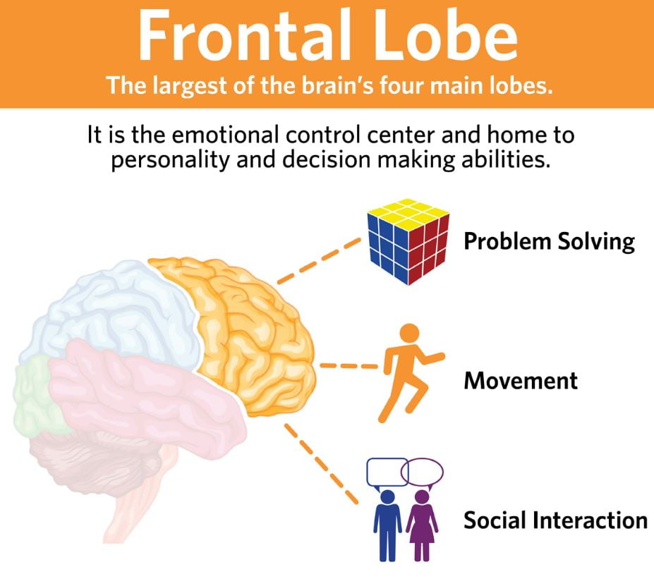
It was with this scale that the attempt to find structures in the brain that would correlate with love began. The only practical conclusion before that was about the fidelity gene, so at first they made a scale like a standard psychological thing - if you want to measure something, do a test. It didn't quite work, so they looked in the brain, and found the nucleus accumbens, which is activated in mice. In fact, they found the same thing. Direct correlation: the higher the score on the love scale, the higher the activation of the nucleus accumbens.
Functional Magnetic Resonance Imaging
Over time, a cohort of scientists emerged to study love through brain imaging, such as MRI. However, this method has its limitations. There are very few ways in which you can show love in an MRI camera, because a person should not move. If a person is in love, then this feeling manifests itself 24/7, and it is impossible to fix a person in an MRI for such an amount of time.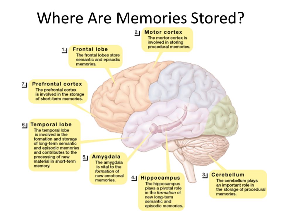 In most experiments, love was measured by looking at photographs of the lover.
The first survey was carried out in 2000. Then the researchers Andreas Bartels and Semir Zeki decided that love manifests itself in the brain if a person is shown a photo of a partner. During the study, 17 subjects were shown photographs of a partner and photographs of a friend whom they have known for the same amount of time and who is similar to their partner in social characteristics. Scientists tried to understand which areas of the brain differ when presented with a photo of a partner and a friend.
The results of the study showed that when a person looks at a lover and realizes that he has a photo of his partner in front of him, the area of \u200b\u200bthe brain responsible for memories is activated in his brain. In addition, it was possible to find out that the insular lobe, which is responsible for emotions, is activated. It turns out that when a person looks at a photo of a partner, he remembers the events associated with him, and these memories have a strong emotional coloring.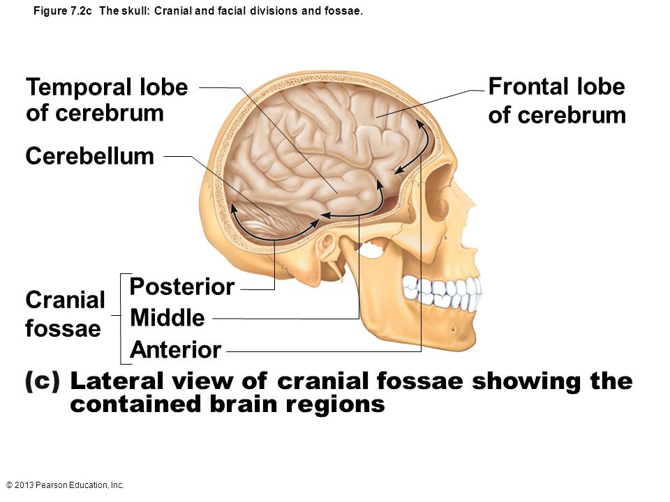
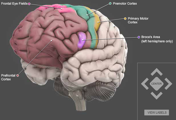
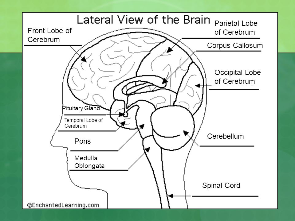
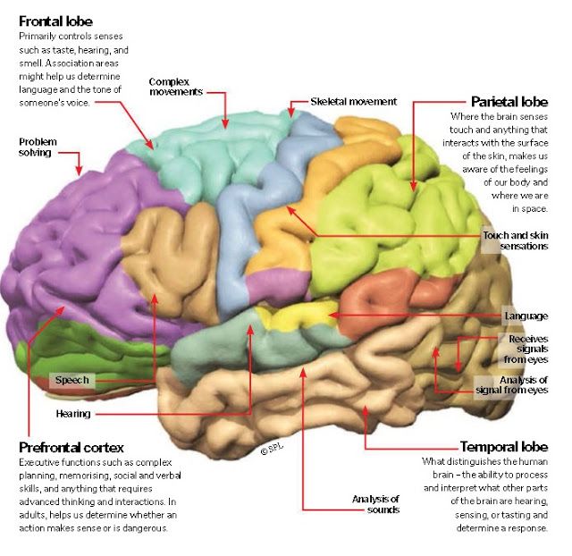
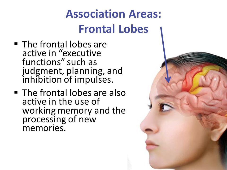
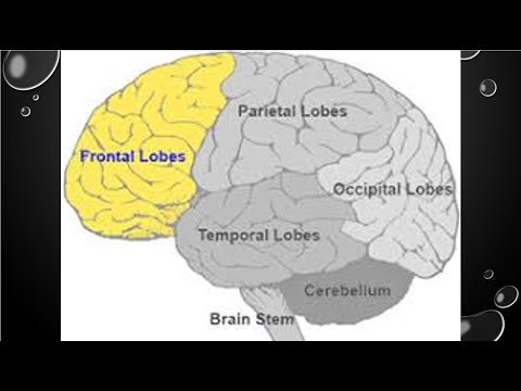
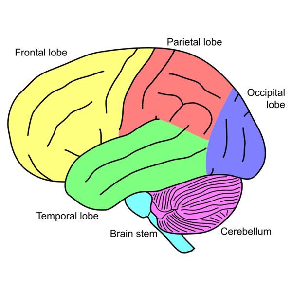
Later scientists decided to compare romantic love with maternal love. There were more subjects in this study. The design of the experiment is similar: the subjects lying in the MRI were shown photographs of their partner and mother, and they tracked what was changing in the brain. Despite the fact that in two types of love, some regions in the brain overlap, there are areas that are characteristic of motherly love. So, the frontal lobes are activated, that is, at the sight of a photograph of the mother, not only the areas responsible for emotional memories are activated, but also the logical components that are associated with the establishment of social relations.
Another study, which also used MRI, showed that when people had a painful tear, when the abandoned person was shown a picture of the initiator of the tear, areas of the brain that are activated when physical pain is felt are activated. In other words, photos of an ex are a serious blow to the one who was dumped (a fact confirmed even scientifically).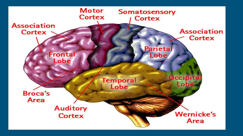 However, MRI is not ideal. Scientists look at the pictures and say that certain areas are activated, which means that the events are equivalent. To make sure that this is not a coincidence, it is necessary to continue to conduct research in this area.
The brain of a man and a woman
On average, women have a thicker corpus callosum and two commissures (constrictions, connections in the brain) - all three brain structures connect the hemispheres with each other. In other words, women have better hemisphere connectivity, so the hemispheres communicate better. The right hemisphere is considered to be responsible for emotions, and speech is controlled by the left for the majority, so it is easier for women to express emotions - what they feel, they transmit to the speech center faster. Another difference between the brain of a man and a woman is that when a person performs a task, the man tries to coordinate the work of the left and right hemispheres, and the woman connects the front and back sections of the brain - analytical and visual-auditory.
The hypothalamus is a structure located in the depths of the brain, it is there that the differences between the male and female brains are clearly visible. If earlier, speaking about love, we talked about the general feeling of affection, which is formed by neurotransmitters and hormones, then we tried to describe love by activation of brain regions at the sight of a lover, then at the level of the hypothalamus, scientists tried to find out which regions are activated during arousal. In women, this is the ventromedial nucleus, an area of the brain that is activated when a woman is ready for sexual intercourse. Men have a middle preoptic region that is responsible for the overall feeling of arousal, but evolution has endowed men with a special INAh4 flare that has the sole function of causing an erection. Women also have such a nucleus, but it is half as large and takes on the functions of neighboring nuclei. It is known that in homosexual men the size of such a nucleus is the same as in women.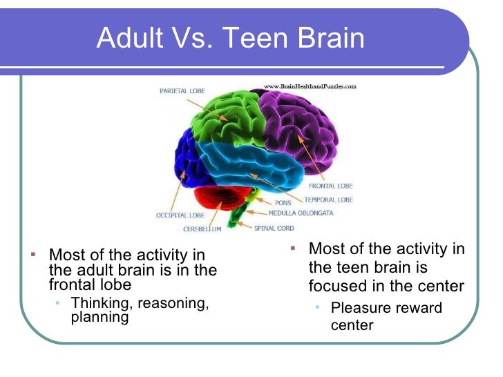 According to this parameter, the brain of homosexuals is similar to the brain of women. Based on this, scientists suggest that homosexuality is a genetically innate phenomenon, since the nuclei are formed genetically.
What happens in the brain of a man and a woman after sex?
Before sex, both sexes have approximately the same situation: the limbic system is activated, which is responsible for emotions, and the areas that are responsible for the analysis of tactile sensations are activated. After an orgasm, different processes occur in the brain of a man and a woman. In men, the activity of the amygdala is sharply reduced, so they become calm and non-aggressive. In women, the activity of the dorsal prefrontal cortex, which is responsible for morality, decreases. Also, in women, the activity of the orbitofrontal cortex, which is responsible for the constant analysis of the environment and the search for dangerous stimuli, decreases.
How brain damage affects human sexual behavior
Most often, changes in human behavior occur when the hypothalamus is damaged, that is, when the frontal and temporal lobes are damaged.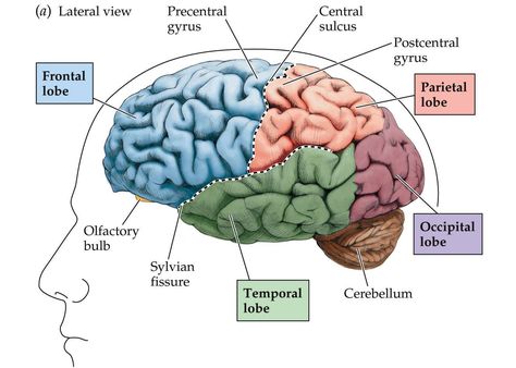 The peculiarity of the brain is that it is closed in the cranium, and if the tumor grows, it presses on neighboring areas. When pressure occurs, certain areas of the brain are activated. If a tumor has grown in the temporal lobe and presses on the hypothalamus, then a person can experience an orgasm just by walking down the street, since the tumor has pressed on the desired nucleus.
There is also a bad scenario for the development of events at the junction of the frontal and temporal lobes. One day, a school teacher realized that he was attracted to the girls he taught. The teacher went to the doctor, and it turned out that a tumor appeared on the border of the frontal and temporal lobes, which led to the manifestation of pedophilic inclinations (while the teacher himself realized that this was wrong). The tumor was removed and the symptoms disappeared, but then the tumor reappeared and a second operation was needed. A tumor in the brain can lead to noticeable external changes in sexual behavior, because those structures that are responsible for sexual arousal and love can be activated both by other people and due to the physical pressure of the tumor that has arisen in the neighboring region.