What scan shows brain activity
Brain PET scan : MedlinePlus Medical Encyclopedia
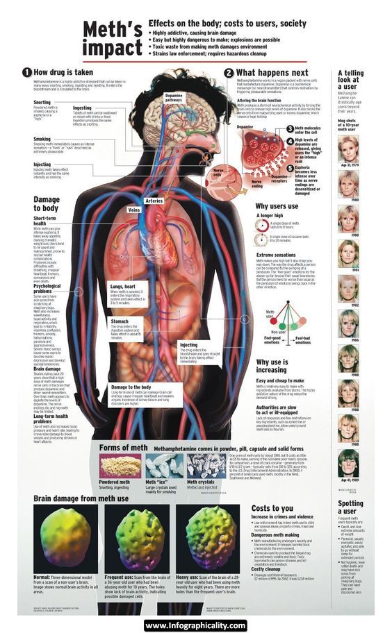
A brain positron emission tomography (PET) scan is an imaging test of the brain. It uses a radioactive substance called a tracer to look for disease or injury in the brain.
A PET scan shows how the brain and its tissues are working. Other imaging tests, such as magnetic resonance imaging (MRI) and computed tomography (CT) scans only reveal the structure of the brain.
A PET scan requires a small amount of radioactive material (tracer). This tracer is given through a vein (IV), usually on the inside of your elbow. Or, you breathe in the radioactive material as a gas.
The tracer travels through your blood and collects in organs and tissues. The tracer helps your health care provider to see certain areas or diseases more clearly.
You wait nearby as the tracer is absorbed by your body. This usually takes about 1 hour.
Then, you lie on a narrow table, which slides into a large tunnel-shaped scanner. The PET scanner detects signals from the tracer. A computer changes the results into 3-D pictures. The images are displayed on a monitor for your provider to read.
You must lie still during the test so that the machine can produce clear images of your brain. You may be asked to read or name letters if your memory is being tested.
The test takes between 30 minutes and 2 hours.
You may be asked not to eat anything for 4 to 6 hours before the scan. You will be able to drink water.
Tell your provider if:
- You are afraid of close spaces (have claustrophobia). You may be given a medicine to help you feel sleepy and less anxious.
- You are pregnant or think you might be pregnant.
- You have any allergies to injected dye (contrast).
- You have taken insulin for diabetes. You will need special preparation.
Always tell your provider about the medicines you are taking, including those bought without a prescription. Sometimes, medicines interfere with the test results.
You may feel a sharp sting when the needle containing the tracer is placed into your vein.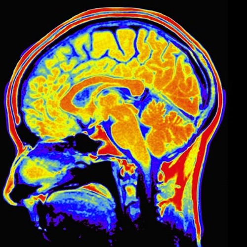
A PET scan causes no pain. The table may be hard or cold, but you can request a blanket or pillow.
An intercom in the room allows you to speak to someone at any time.
There is no recovery time unless you were given a medicine to relax.
After the test, drink a lot of fluids to flush the tracer out of your body.
A PET scan can show the size, shape, and function of the brain, so your doctor can make sure it is working as well as it should. It is most often used when other tests, such as MRI scan or CT scan, do not provide enough information.
This test can be used to:
- Diagnose cancer
- Prepare for epilepsy surgery
- Help diagnose dementia if other tests and exams do not provide enough information
- Tell the difference between Parkinson disease and other movement disorders
Several PET scans may be taken to determine how well you are responding to treatment for cancer or another illness.
There are no problems detected in the size, shape, or function of the brain.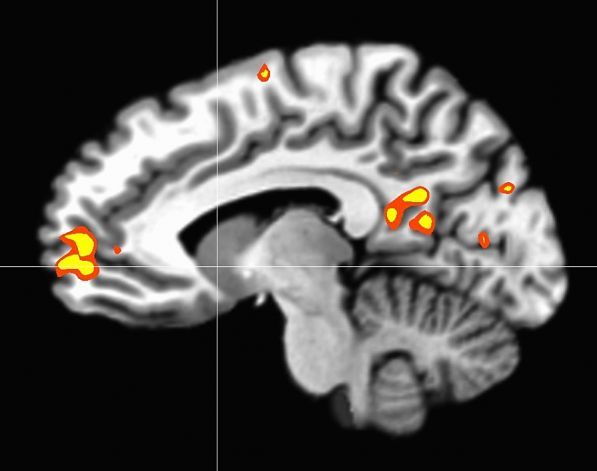 There are no areas in which the tracer has abnormally collected.
Abnormal results may be due to:
- Alzheimer disease or dementia
- Brain tumor or spread of cancer from another body area to the brain
- Epilepsy, and may identify where the seizures start in your brain
- Movement disorders (such as Parkinson disease)
The amount of radiation used in a PET scan is low. It is about the same amount of radiation as in most CT scans. Also, the radiation does not last for long in your body.
Women who are pregnant or are breastfeeding should let their provider know before having this test. Infants and babies developing in the womb are more sensitive to the effects of radiation because their organs are still growing.
It is possible, though very unlikely, to have an allergic reaction to the radioactive substance. Some people have pain, redness, or swelling at the injection site.
It is possible to have false results on a PET scan. Blood sugar or insulin levels may affect the test results in people with diabetes.
PET scans may be done along with a CT scan. This combination scan is called a PET/CT.
Brain positron emission tomography; PET scan - brain
Chernecky CC, Berger BJ. Positron emission tomography (PET) - diagnostic. In: Chernecky CC, Berger BJ, eds. Laboratory Tests and Diagnostic Procedures. 6th ed. St Louis, MO: Elsevier Saunders; 2013:892-894.
Glaudemans AWJM, Israel O, Slart RHJA, Ben-Haim S. Vascular PET/CT and SPECT/CT. In: Sidawy AN, Perler BA, eds. Rutherford's Vascular Surgery and Endovascular Therapy. 9th ed. Philadelphia, PA: Elsevier; 2019:chap 29.
Hutton BF, Segerman D, Miles KA. Radionuclide and hybrid imaging. In: Adam A, Dixon AK, Gillard JH, Schaefer-Prokop CM, eds. Grainger & Allison's Diagnostic Radiology: A Textbook of Medical Imaging. 6th ed. Philadelphia, PA: Elsevier Churchill Livingstone; 2015:chap 6.
Jankovic J, Mazziotta JC, Newman NJ, Pomeroy SL. Investigations in the diagnosis and management of neurological disease. In: Jankovic J, Mazziotta JC, Pomeroy SL, Newman NJ, eds. Bradley and Daroff's Neurology in Clinical Practice
. 8th ed. Philadelphia, PA: Elsevier; 2022:chap 34.
Updated by: Evelyn O. Berman, MD, Assistant Professor of Neurology and Pediatrics at University of Rochester, Rochester, NY. Review provided by VeriMed Healthcare Network. Also reviewed by David Zieve, MD, MHA, Medical Director, Brenda Conaway, Editorial Director, and the A.D.A.M. Editorial team.
Brain scans – MRI scan – CT scan – BHF
Brain scans produce detailed images of the brain. They can be used to help doctors detect and diagnose conditions, such as tumours, causes of a stroke or vascular dementia.
The two most common types of brain scans are:
• Magnetic Resonance Imaging (MRI scans)
• Computerised Tomography (CT scans)
Brain MRI scans
MRI scans use magnetic fields and radio waves to produce images. An MRI scan of the head produces a detailed image of the brain and brain stem.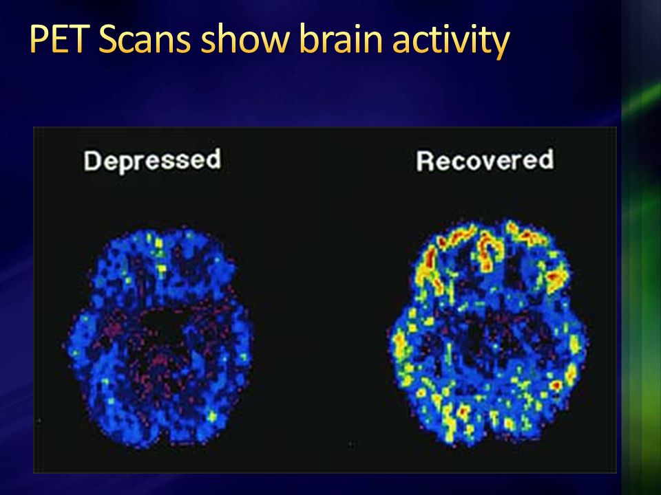
Why am I having a brain MRI?
From your brain MRI scan, doctors can understand whether you’ve had a stroke or have vascular dementia, or both. It may also be used to investigate whether you have any other conditions, such as cancer.
An MRI will be used to investigate why you’re experiencing symptoms. If you have any questions or concerns, you should talk to your doctor.
Before a brain MRI scan
You usually won't need to prepare for a brain MRI scan, but as it involves magnets you will need to remove anything that contains metal, including jewellery and hairpins.
Before the scan, you may be injected with a dye called contrast, through an a cannula which is placed into a vein in the arm. This allows the MRI scanner to see your brain in detail.
During a brain MRI scan
During a brain MRI scan:
- you'll need to keep still during the scan, so that the images come out clear
- you'll be asked to lie on a bed that’s slowly moved head first into an MRI scanner
- the scanner will make loud tapping sounds
- you’ll be given earplugs or headphones so you can listen to music to distract you from the noise.

The scan may last between 15 and 90 minutes, depending on how many images of the brain are needed.
Are brain MRI scans safe?
An MRI scan is painless and safe. You may wish to tell the technician doing the scan if you have a fear of confined spaces, so that they can try to make you as comfortable as possible.
You may not be able to have an MRI scan if you have a pacemaker or implantable defibrillator, but this will have been discussed with you beforehand. You will need to sign a checklist before you have it done to ensure it is safe for you to have the scan.
- Find out more about MRI scans for the heart, also known as cardiac MRI scans.
Brain CT scans
A CT scan uses X-rays to produce images, unlike an MRI scan which uses magnetic fields and radio waves.
Why am I having a brain CT scan?
A CT scan can detect conditions of the brain, like stroke and vascular dementia. The images produced by a CT scan provide detailed information about brain tissue and brain structures.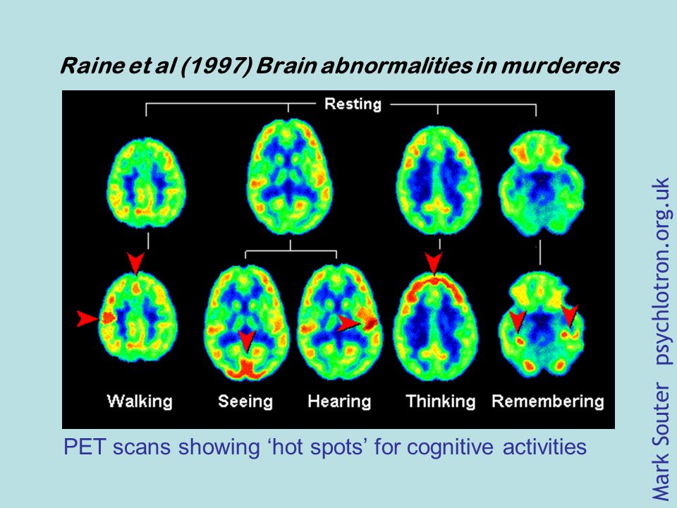
Preparing for a brain CT scan
You will likely receive a letter confirming your CT scan, which will tell you anything you need to do beforehand. Right before the scan, you will need to remove anything that contains metal, including jewellery and hairpins. You may also be given a contrast solution that helps to improve the quality of the images.What happens during a brain CT scan
During a CT scan,
- you will need to lie on a bed that’s slowly moved into a CT scanner head first
- the scanner will rotate around your head.
It’s important to keep as still as possible during the scan, so that the images come out clear. CT scans are quicker than MRI scans, usually lasting about 10 minutes.
Are brain CT scans safe?
CT scans expose you to X-rays, but the scanners are designed so that you’re not exposed to very high levels of radiation. Your doctor will always weigh up the benefits and risks of a CT scan before advising you to have one.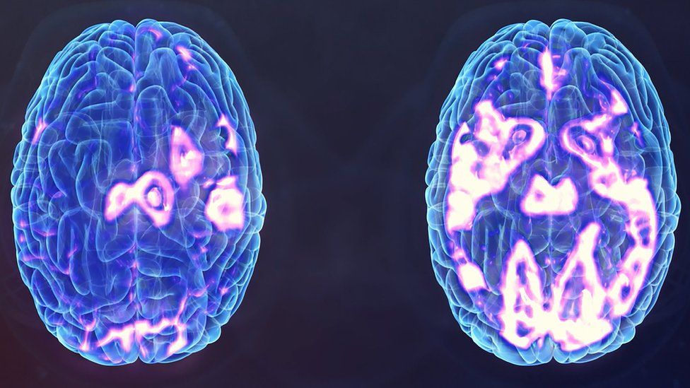
- For more information on brain CT scans, read our Q&A with Professor Joanna Wardlaw, What is a CT scan of the brain?
EEG (electroencephalography) - what it shows, how it is done, preparation
What it is
What it shows
How it is done
Indications
Contraindications
Preparation
Electroencephalography (EEG) is a method of studying the electrical activity of the brain by placing electrodes in certain areas on the surface of the head.
What is the electrical activity of the brain
The brain consists of nerve cells - neurons that have the ability to transmit electrical impulses "along the chain". Different parts of the brain react to various external stimuli - within these areas, neurons transmit a single impulse. In addition, under certain conditions, the impulses can weaken or strengthen each other.
The electrical impulses arising in the brain are able to catch the electroencephalograph. It consists of electrodes attached to a computer. Electrodes attached to the patient's head pick up the pulses and transmit them to a computer for interpretation and display. On paper, impulses appear as waves. Waves differ in characteristics (frequency and amplitude) and are divided into alpha, beta, delta, theta and mu waves.
What does the EEG show
An electroencephalogram allows a specialist to see signs of various disorders of the brain and assess their nature. For example, using an EEG, you can recognize:
- epileptic activity in various parts of the brain;
- possible causes of panic attacks and loss of consciousness;
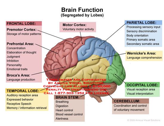
- in which lobes of the brain are pathological foci located;
- how the electrical activity of the brain changes before attacks.
In addition, using the EEG, you can find out what causes neurological problems - functional disorders or an organic lesion, as well as evaluate the effectiveness of therapy and rehabilitation (in this case, the EEG is taken before the start of treatment, and then - during or after a course of medications).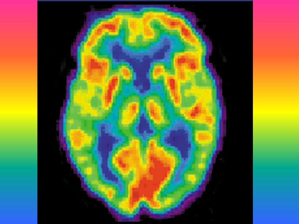
How an EEG is performed
A cap with electrodes attached to it is put on the patient's head. The doctor examines the electrical activity of the patient's brain at rest, asks him to blink to take into account errors in blinking, and then additionally influences the patient by asking him to breathe deeply (hyperventilation) and studying his reaction to flashes of light (photostimulation).
EEG indications
- Past traumatic brain injury or neurosurgical interventions.
- Circulatory disorders in the brain (for example, strokes).
- Suspected brain tumor.
- History of seizures or syncope.
- Frequent bouts of dizziness and headaches.
- Various sleep disorders (insomnia, frequent nocturnal awakenings, somnambulism).
- Meningitis, encephalitis and other diseases of the meninges.
- Delayed speech and psychomotor development, stuttering.
- Tics in children.
- Cerebral palsy.
EEG contraindications
There are no clear contraindications to the procedure. In some cases, the procedure may be difficult due to open wounds and injuries that interfere with the connection of electrodes, as well as in young children and people with certain mental disorders, since it is difficult for them to maintain the calmness necessary for conducting an examination and accurately interpreting the result.
Preparation for EEG
- Do not take anticonvulsants, sedatives and invigorating drugs 3-4 days before the study, also limit coffee and strong tea, energy drinks and chocolate.
- Before the procedure, the head should be well washed and cleaned of styling products, the hair should be dried.
- Eat well a couple of hours before the test to prevent hunger and a drop in blood sugar, which can skew the results of the test.
- If EEG is planned to be performed while asleep (usually required for epilepsy), the night before the study should be sleepless.

The author of the article:
Zorkina Lyubov Alekseevna
Doctor of functional diagnostics, ultrasound
work experience 9 years
reviews leave feedback
Clinic
m. Red Gate
Services
- Name
- Electroencephalography (EEG)2900
Health articles
All articlesAllergistGastroenterologistHematologistGynecologistDermatologistImmunologistInfectionistCardiologistCosmetologistENT doctor (otolaryngologist)MammologistMassageNeurologistNephrologistOzone therapyOncologistOphthalmologistProctologistPsychotherapistPulmonologistRheumatologistTherapistTraumatologistTrichologistUltrasound (ultrasound examination)UrologistPhysiotherapistPhlebologistSurgeonFunctional diagnostics and Energist 905 years. Red Gates. AvtozavodskayaPharmacy. Glades. Sukharevskaya. st. Academician Yangelam. Frunzenskaya ZelenogradDmitrieva Olga Nikolaevna
Chief physician of "Polyclinika. ru" on Frunzenskaya, neurologist, ENMG specialist
reviews
Clinic
m. Frunzenskaya
Stolbova Tatyana Sergeevna
Chief physician of "Polyclinika.ru" on the street. 1905, therapist, gastroenterologist
reviews
Clinic
m. Street 1905 Goda
Yuliy Sergeevich Gromov
Head physician of "Polyclinic.ru" on Sukharevskaya, surgeon
reviews
Clinic
m. Sukharevskaya
Agarkova Elena Valentinovna
Head physician "Polyclinika.ru" in Zelenograd, gastroenterologist
reviews
Clinic
Zelenograd
Aydinov Gennady Ivanovich
Chief Physician "Polyclinic. ru" Academician Yangel, therapist
reviews
Clinic
m. st. Academician Yangel
Godulyan Aleksey Viktorovich
chief physician of "Polyclinika.ru" at Krasnye Vorota, endocrinologist, KMN
reviews
Clinic
m. Red Gate
Kupriyanova Olga Sergeevna
Head doctor "Polyclinika.ru" on Avtozavodskaya, general practitioner
reviews
Clinic
m. Avtozavodskaya
Sumina Evgenia Yuryevna
Chief physician of "Polyanka.ru" in Polyanka, neurologist
reviews
Clinic
m.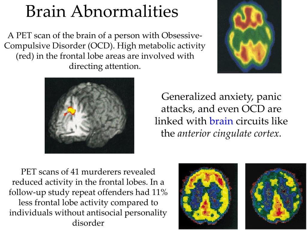 Polyanka
Tolstikova Anna Pavlovna
Chief physician of "Polyclinic.ru" on Taganskaya, general practitioner
reviews
Clinic
m. Taganskaya
Shishkina Svetlana Valerievna
Medical Director
reviews
Clinic
01/653
Brain scan helps to distinguish a deliberate crime from a crime committed through negligence In court, as a rule, premeditated crimes and those committed in a state of passion or negligence have a different legal assessment. True, it is extremely difficult to determine the state of a person during the commission of a crime after everything has already happened. Understanding this, many criminals who have committed serious crimes, such as murder, try to present it as being committed in a state of passion or thoughtlessness.
 Malefactors know - it will be possible to prove, for example, manslaughter, the punishment will be milder than if the judge considers the crime to be intentional, and even planned in advance.
 Modern technology can help the court distinguish truth from deceit, cold-blooded murder from unconsciousness. A recent study published in PNAS (Proceedings of the National Academy of Sciences) shows that the brain activity of criminals who commit crimes with a clear mind and solid memory differs from that of people who commit a crime unknowingly. A study by a team from the Salk Institute for Biological Research found that brain scans of suspects can help distinguish between the two.
Functional magnetic resonance imaging is used for this operation. This is the name of a type of magnetic resonance imaging, which is performed to measure hemodynamic reactions (changes in blood flow) caused by neuronal activity in the brain or spinal cord. The method is based on the fact that cerebral blood flow and neuronal activity are interconnected.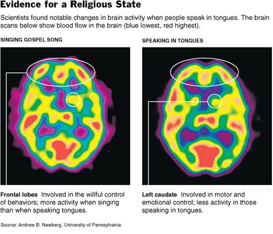 When an area of the brain is active, blood flow to that area also increases.
Scan results are fed to a self-learning computer system with a specialized neural network. To implement their project, scientists invited several volunteers who agreed to scan and process its results. Only these were not hardened criminals at all - murderers, thieves and swindlers. No, the experiment involved ordinary people who were asked to commit crime models, so to speak.
For example, briefcases with certain goods were placed on the table. The participants of the experiment were asked to choose one of the briefcases, which then had to be carried through a guarded point with a guard and a scanner (the point and the guard were also provided by the experimenters, nothing illegal). Moreover, they were given to know that “smuggled goods” were invested somewhere. Representatives of the control group did not know which portfolio contains "smuggling". But the participants in the experiment from the other group were aware of this - they were warned about "smuggling", but the choice was limited to just one portfolio.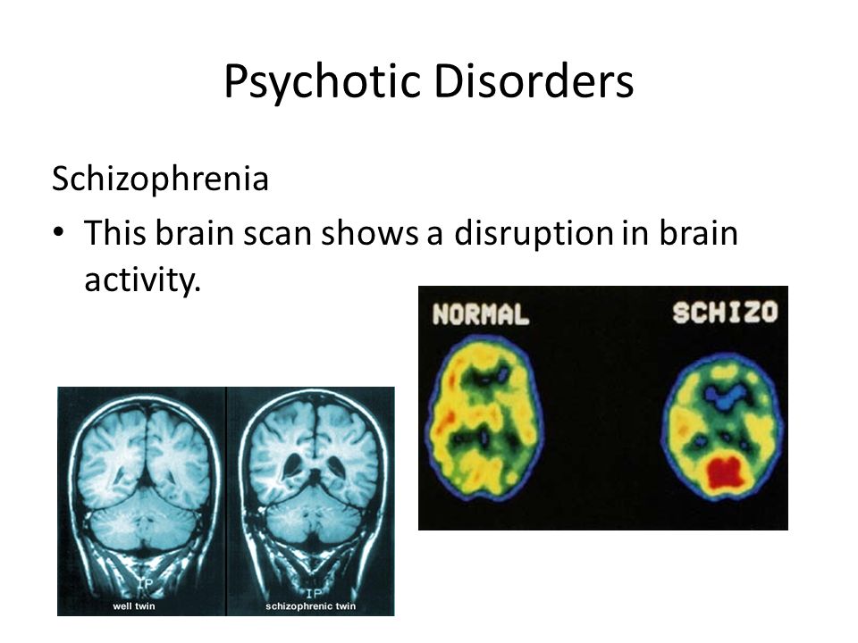 Thus, people from the first group were considered as "criminals" who committed an unconscious crime. People from the second group were considered as criminals who knew perfectly well what they were getting into. Despite some simplicity and naivety of this experiment, scientists considered that the brain activity of volunteers in the described situation is comparable to the brain activity of real criminals.
The scientists who conducted the experiment scanned the brain activity of all participants using functional magnetic resonance imaging with additional magnetic resonance imaging data. This is already a way to obtain tomographic medical images for the study of internal organs and tissues using the phenomenon of nuclear magnetic resonance. It is based on measuring the electromagnetic response of atomic nuclei, most often the nuclei of hydrogen atoms, namely, on their excitation by a certain combination of electromagnetic waves in a constant magnetic field of high intensity.
The computer system, after analyzing the tomography data, showed that the "criminals" who violated, realizing the presence of contraband, activated other parts of the brain than those volunteers who walked through the checkpoint, not knowing if everything was in order with things in their portfolio.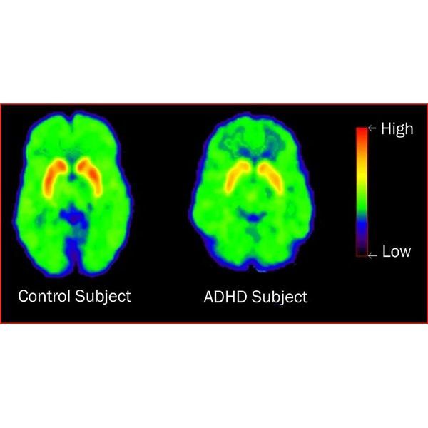 In the first case, the analysis showed the activity of the middle prefrontal cortex of the brain. This, according to modern scientists, is the area that is responsible for the self-awareness of a person. She is also responsible for the behavior of a person who expects a reward or punishment. Also in this case, the temporo-parietal region became active, starting to work when a person utters a deliberate lie.
Those volunteers who were not aware of what was in the briefcase activated the occipital cortex, which is mainly responsible for processing visual information.
The results of the study are still preliminary. However, it is likely that criminals who consciously engage in a dark business analyze the risks, assessing the situation from different angles. Those who committed the crime unknowingly think differently.
The authors of the experiment also tested two more scenarios. In the first case, those who knew about the smuggling in the briefcase were informed of the impending inspection by the security guard.


