Intermittent explosive disorder icd 10


2023 ICD-10-CM Diagnosis Code F63.81: Intermittent explosive disorder

  1. ICD-10-CM Codes
  2. F01-F99
  3. F60-F69
  4. F63-
  5. 2023 ICD-10-CM Diagnosis Code F63.81

Intermittent explosive disorder

    2016 2017 2018 2019 2020 2021 2022 2023 Billable/Specific Code
  • F63.81 is a billable/specific ICD-10-CM code that can be used to indicate a diagnosis for reimbursement purposes.
  • The 2023 edition of ICD-10-CM F63.81 became effective on October 1, 2022.
  • This is the American ICD-10-CM version of F63.81 - other international versions of ICD-10 F63.81 may differ.

The following code(s) above F63. 81 contain annotation back-references

Annotation Back-References

In this context, annotation back-references refer to codes that contain:

  • Applicable To annotations, or
  • Code Also annotations, or
  • Code First annotations, or
  • Excludes1 annotations, or
  • Excludes2 annotations, or
  • Includes annotations, or
  • Note annotations, or
  • Use Additional annotations

that may be applicable to F63.81:

  • F01-F99

    2023 ICD-10-CM Range F01-F99

    Mental, Behavioral and Neurodevelopmental disorders

    Includes

    • disorders of psychological development

    Type 2 Excludes

    • symptoms, signs and abnormal clinical laboratory findings, not elsewhere classified (R00-R99)
    Mental, Behavioral and Neurodevelopmental disorders
  • F63

    ICD-10-CM Diagnosis Code F63

    Impulse disorders

      2016 2017 2018 2019 2020 2021 2022 2023 Non-Billable/Non-Specific Code

    Type 2 Excludes

    • habitual excessive use of alcohol or psychoactive substances (F10-F19)
    • impulse disorders involving sexual behavior (F65. -)
    Impulse disorders

Approximate Synonyms

  • Explosive disorder, intermittent
  • Explosive disorder, isolated
  • Isolated explosive disorder

Clinical Information

  • A disorder characterized by recurrent episodes of serious assaultive acts or destruction of property due to a failure to resist aggressive impulses; the degree of aggression during these episodes is grossly out of proportion to any psychosocial provocation. The aggressive episodes are not etiologically linked to another mental disorder, a general medical condition, or substance use.

ICD-10-CM F63.81 is grouped within Diagnostic Related Group(s) (MS-DRG v40.0):

  • 883 Disorders of personality and impulse control

Convert F63. 81 to ICD-9-CM

Code History

  • 2016 (effective 10/1/2015): New code (first year of non-draft ICD-10-CM)
  • 2017 (effective 10/1/2016): No change
  • 2018 (effective 10/1/2017): No change
  • 2019 (effective 10/1/2018): No change
  • 2020 (effective 10/1/2019): No change
  • 2021 (effective 10/1/2020): No change
  • 2022 (effective 10/1/2021): No change
  • 2023 (effective 10/1/2022): No change

Diagnosis Index entries containing back-references to F63.81:

  • Disorder (of) - see also Disease
    • intermittent explosive F63.81

ICD-10-CM Codes Adjacent To F63. 81

F60.8 Other specific personality disorders

F60.81 Narcissistic personality disorder

F60.89 Other specific personality disorders

F60.9 Personality disorder, unspecified

F63 Impulse disorders

F63. 0 Pathological gambling

F63.1 Pyromania

F63.2 Kleptomania

F63.3 Trichotillomania

F63.8 Other impulse disorders

F63.81 Intermittent explosive disorder

F63. 89 Other impulse disorders

F63.9 Impulse disorder, unspecified

F64 Gender identity disorders

F64.0 Transsexualism

F64.1 Dual role transvestism

F64.2 Gender identity disorder of childhood

F64. 8 Other gender identity disorders

F64.9 Gender identity disorder, unspecified

F65 Paraphilias

F65.0 Fetishism

Reimbursement claims with a date of service on or after October 1, 2015 require the use of ICD-10-CM codes.

ICD-9-CM Diagnosis Code 312.34 : Intermittent explosive disorder

Home > 2015 ICD-9-CM Diagnosis Codes > Mental Disorders 290-319 > Neurotic Disorders, Personality Disorders, And Other Nonpsychotic Mental Disorders 300-316 > Disturbance of conduct not elsewhere classified 312-

2015 ICD-9-CM Diagnosis Code 312. 34

Intermittent explosive disorder

  • 2015
  • Billable Thru Sept 30/2015
  • Non-Billable On/After Oct 1/2015

  • ICD-9-CM 312.34 is a billable medical code that can be used to indicate a diagnosis on a reimbursement claim, however, 312.34 should only be used for claims with a date of service on or before September 30, 2015. For claims with a date of service on or after October 1, 2015, use an equivalent ICD-10-CM code (or codes).

Convert to ICD-10-CM: 312.34 converts approximately to:

  • 2015/16 ICD-10-CM F63.81 Intermittent explosive disorder

Approximate Synonyms

  • Explosive disorder, intermittent

Clinical Information

  • A disorder characterized by recurrent episodes of serious assaultive acts or destruction of property due to a failure to resist aggressive impulses; the degree of aggression during these episodes is grossly out of proportion to any psychosocial provocation. The aggressive episodes are not etiologically linked to another mental disorder, a general medical condition, or substance use

ICD-9-CM Volume 2 Index entries containing back-references to 312.34:

  • Disorder - see also Disease
    • explosive
      • intermittent 312.34
      • isolated 312.35
    • intermittent explosive 312.34
  • Disturbance - see also Disease
    • conduct 312.9
      • adjustment reaction 309.3
      • adolescent onset type 312.82
      • childhood onset type 312.81
      • compulsive 312.30
        • intermittent explosive disorder 312.34
        • isolated explosive disorder 312.35
        • kleptomania 312.32
        • pathological gambling 312.31
        • pyromania 312.33
      • hyperkinetic 314.2
      • intermittent explosive 312.34
      • isolated explosive 312.35
      • mixed with emotions 312.4
      • socialized (type) 312.20
        • aggressive 312.23
        • unaggressive 312. 21
      • specified type NEC 312.89
      • undersocialized, unsocialized
        • aggressive (type) 312.0
        • unaggressive (type) 312.1
 312.33 ICD9Data.com 312.35 
  • 2015/Index · 2014/Index · 2013/Index
  • 2012/Index · 2011/Index · 2010/Index
  • 2009/Index · 2008/Index · 2007/Index
  • About · Privacy Policy · Disclaimer
  • 2016 ICD-10-CM Diagnosis Codes · Index
  • Convert ICD-9-CM <-> 2016 ICD-10-CM
  • 2016 ICD-10-PCS Procedure Codes
  • 2016 · 2015 · 2014 · 2013 · 2012
  • 2011 · 2010 · 2009 · 2008

Intermittent Explosive Disorder - frwiki.wiki

Intermittent temper disorder ( TEI ) is a behavioral disorder characterized by extreme expressions of anger, often to the point of violence, disproportionate to what is happening (see also Seb Buje's definition). It is currently classified in the Diagnostic and Statistical Manual of Mental Disorders as habit and impulse disorder. TEI belongs to axis I of the habit and impulse disorders listed in the DSM-IV-TR, which include kleptomania, pyromania, pathological gambling, and others. Impulsive aggression is not intentional and is defined by a disproportionate response to every perceived response. Some people report physiological changes (eg, tension, depression).

A 2006 study shows that the prevalence is much higher than previously thought.

Prevalence is higher in men than in women. The disorder itself is not easily characterized and often comorbid with other mood disorders, especially bipolar disorder. People diagnosed with TEI report that their temper tantrums are short-lived (lasting an hour or less) and are accompanied by a variety of symptoms (sweating, heart palpitations). Violent acts are often reported accompanied by a sense of relief or even in some cases pleasure, but subsequently accompanied by remorse. Then the person is in a phase of complete loss of control. Moments of extreme anger followed a hidden headache, severe chest pain (sharp burning) and eye pain (or even red eyes). Which sometimes leads to death.

CV

  • 1 Diagnosis
  • 2 Treatment
  • 3 links
  • 4 External link

Diagnostic

Diagnostic criteria for DSM-IV include: inability to resist aggressive urges, level of aggression expressed during a highly disproportionate episode in the face of a provocation or source of psychosocial stress, and, as previously explained, a diagnosis is made when other mental disorders that may be harmful (eg, antisocial personality disorder, borderline personality). At a distance, acts of aggression cannot be caused by a common disease, for example. head injuries, Alzheimer's disease, etc. as a result of long-term drug use or drug abuse. The diagnosis is made when the emotional and behavioral symptoms meet the criteria specified in the DSM-IV.

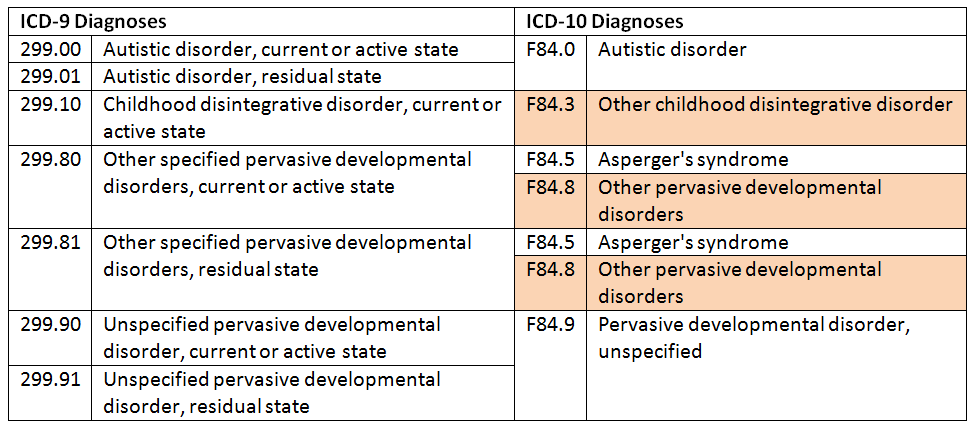
The DSM-IV-TR is very selective in its definitions of intermittent explosive disorder, which essentially defines the exclusion of other conditions. Diagnosis requires:

  1. Several episodes of impulsive behavior that could result in very serious damage to another person or property.
  2. The level of aggression is grossly disproportionate to the circumstances or the provocation.
  3. Episodic violence cannot be caused by another physical or mental illness.

Care

Treatment, which requires, inter alia, cognitive behavioral therapy and psychotropic drugs, has shown temporary success. Therapy can help the patient recognize their impulsivity and control it. Patients with TEI are prescribed several drugs.

Of the tricyclic antidepressants and selective serotonin reuptake inhibitors such as fluoxetine, fluvoxamine and sertraline appear to relieve some psychopathological symptoms.

Recommendations

  1. Diagnostic and Statistical Manual of Mental Disorders: DSM-IV , Washington, DC, American Psychiatric Association , 943 p. (ISBN 0-89042-025-4) , 663–7
  2. (in) McElroy S. L., Recognition and treatment of intermittent explosive disorder DSM-IV , theft. 60 Appendix 15, , 12–6 p. (PMID 10418808)
  3. (in) Boyd, Mary Ann, The Psychiatric Nurse: Modern Practice , Philadelphia, Wolters Kluwer Health / Lippincott Williams & Wilkins, , 820–1 pp. (ISBN 0-7817-9169-3)
  4. a b and c (ru) McElroy SL, Soutullo CA, Beckman DA, Taylor P, Keck PE, DSM-IV intermittent explosive disorder: report of 27 cases , T. 59, , 203–10; quiz 211 p. (PMID 95
  5. , DOI 10.4088 / JCP.v59n0411)
  6. (in) McCloskey, MS, Noblett, KL, Deffenbacher, JL Gollan, JK Coccaro, EF (2008) Cognitive behavioral therapy for intermittent explosive disorder: a pilot randomized clinical trial. 76(5), 876-886.

External link

  • (en) « Impulsive Disorder Management Clinic at the University of Minnesota » ( Archive • Wikiwix • Archive.is • Google • What to do? )

Mental and behavioral disorders (ICD-10: F00-F99)

( F00-F09 ) Organic mental disorders, including symptomatic disorders
Dementia
  • Vascular dementia
  • Huntington's chorea
  • Leukoaraiosis
  • Alzheimer's disease
  • Creutzfeldt-Jakob disease
  • Parkinson's disease
  • Pick's disease
Other
  • Amnesia
  • Brad
  • Other
    • Postconcussion syndrome
    • Mild cognitive impairment
    • Frontal syndrome
( F10-F19 ) Substance use-related mental and behavioral disorders
  • Acute poisoning
    • Ownership status
    • Trance state
    • Acute alcohol intoxication
    • Alcohol poisoning
    • Bad trips
  • Abuse
  • addiction syndrome
    • Chronic alcoholism
    • Dipsomania
    • Substance dependence
  • Withdrawal syndrome
  • Withdrawal syndrome with delirium
  • Psychotic disorder
    • Alcoholic hallucinosis
  • amnesic syndrome
    • Wernicke-Korsakoff syndrome
( F20-F29 ) Schizophrenia, schizotype and delirium
  • Schizophrenia
    • Paranoid schizophrenia
    • Hebephrenic schizophrenia
    • Catatonic schizophrenia
    • Schizophreniform disorder
  • Schizotypal disorder
  • delusional disorder
    • Paranoia
    • Paraphrenia
    • Paranoid psychosis
  • Picky
  • Oneirophrenia
  • Brief reactive psychosis
  • Induced delusional disorder
  • Schizoaffective disorders
  • Chronic hallucinatory psychosis
  • Non-organic psychosis, unspecified
( F30-F39 ) Mood disorders
  • Mania
    • Hypomania
  • Bipolar disorder
    • Bipolar disorder not specified
  • Depression
    • Psychotic depression
    • Atypical depression
    • Masked Depression
    • Seasonal depression
    • Depressed
  • Cyclothymia
  • Dysthymia
  • Brief recurrent depression
  • Premenstrual dysphoric disorder
( F40-F48 ) Neurotic disorders, stress related disorders and somatoform disorders
Anxiety disorders
  • Phobia
    • Agoraphobia
    • Social phobia (anthropophobia)
    • Specific phobia (acrophobia, claustrophobia, animal phobia)
  • panic disorder
    • Panic attack
  • Generalized alarm
Stress and adaptation
  • Acute stress reaction
  • Post-traumatic stress disorder
  • Adjustment disorder
    • Hospitalism
    • Culture shock
Somatoform disorder
  • Somatization
  • Hypochondriacal disorder
    • Dysmorphophobia
    • Hypochondria
    • Nosophobia
  • Psychalgia
Dissociative (conversion) disorders
  • Dissociative Fugue
  • Dissociative amnesia
  • Other
    • Ganser syndrome
    • Dissociative identity disorder
Other
  • Obsessive Compulsive Disorder (OCD)
  • Other
    • Neurasthenia
    • Syndrome of depersonalization - derealization
    • Psychasthenia
    • Relief syndrome
( F50-F59 ) Behavior related to physiological disorders and physical factors
Eating disorders
  • Anorexia nervosa
    • Male anorexia nervosa
  • Bulimia
  • Psychogenic overeating
  • Psychogenic vomiting
  • Psychogenic loss of appetite
  • Adult pike
  • Non-specific eating disorders
Non-organic sleep disorders
  • Insomnia of inorganic nature
  • Nonorganic hypersomnia
  • Irregular rhythm of sleep and wakefulness
  • Somnambulism
  • Night terror
  • Nightmare
  • Inorganic parasomnia
Sexual dysfunction
  • Lack or loss of sexual desire
  • Sexual impotence
  • Anorgasmia
  • Premature ejaculation
  • Psychogenic vaginismus
  • Psychogenic dyspareunia
  • Hypersexuality
Other
  • Perinatal depression
  • Postpartum psychosis
( F60-F69 ) Adult personality and behavior disorders
Personality problems
  • paranoid
  • schizoid
  • dissocial
  • border
  • theatrical
  • anancastic
  • avoidant
  • dependent
  • Other
    • narcissistic
    • passive-aggressive
    • megalomania
    • mythomania
Gender identity and preferences
  • gender dysphoria
    • Gender dysphoria
    • Eonism, transvestism, bivalent transvestism
    • Gender dysphoria in children
  • Psychological and behavioral disorders related to sexual development and orientation
    • Puberty disorder
    • Sexual arousal disorder
    • Egodystonic sexual orientation
    • Sexual problems in relationships
  • Disorders of sexual preference
    • Fetishism
    • Fetish Transvestite
    • Exhibitionism
    • Voyeurism
    • Pedophilia
    • Sadomasochism
    • Necrophilia
    • Zoophilia
Other
  • Disorder of habits and desires
    • Pathological gambling
    • Kleptomania
    • Pyromania
    • Trichotillomania
    • Intermittent explosive disorder
  • Pathomimia
    • Munchausen syndrome
(F70-F99) Mental disorders diagnosed in childhood
( F70-F79 )
Mental retardation
  • Easy (IQ 50 to 69)
  • Medium (IQ 35 to 49)
  • Severe (IQ 20 to 34)
  • Deep (IQ below 20)
( F80-F89 )
Developmental disorders
  • Speech and language disorders
    • Dyslalia
    • Dysphasia
    • Receptive aphasia
    • Aphasia (Landau and Kleffner syndrome)
    • Lisp
  • Failure to study at school
    • Reading disorder (developmental dyslexia)
    • Dysgraphia
    • Agraphia
    • Acalculia
    • Dyscalculia (Gerstmann syndrome)
    • Dysortography
  • Dyspraxia
  • General Developmental Disorders
    • Childhood autism
    • Atypical autism
    • Rett syndrome
    • Childhood disintegrative disorder
    • Asperger's Syndrome
    • Unspecified disorder
( F90-F98 )
Emotional and behavioral disorders
  • Hyperkinetic disorders
  • Conduct disorders
    • Oppositional defiant disorder
  • Emotional disorders
    • Separation anxiety
    • Childhood phobic anxiety disorder
    • Social anxiety
    • Anxiety
    • Identity disorder
  • Selective mutism
  • Reactive attachment disorder in childhood
  • Attachment disorder
  • Tiki
    • Gilles de la Tourette disease
  • Enuresis
  • Encoprese
  • Children's mericism
  • Infant and child pick
  • Stuttering
  • Squirting (Hurry Tongue)
  • Onychophagia
  • excessive masturbation

Traumatology and orthopedic department №10

8 (3522) 45-33-57, 45-43-06

Block B, 2nd floor ensure the transition of a patient with an axial skeletal pathology, i.e. diseases of the spine, spinal cord, deformities of the chest and pelvis to a qualitatively higher level of support and movement.

History and background of creation:

Traumatology and orthopedic department No. 10 was organized in 2015 on the basis of the department of vertebrology and neurosurgery.

Scientific group and department of vertebrology and neurosurgery was organized in 1993. Khudyaev Alexander Timofeevich headed the department and clinical group. Under the leadership of A.T. Khudyaev, the use of an external transpedicular fixation apparatus was started in the treatment of patients with traumatic injuries of the spine, scoliotic and kyphotic deformities, and spondylolisthesis.

The first experimental work on the spine refers to the end of 1974. 
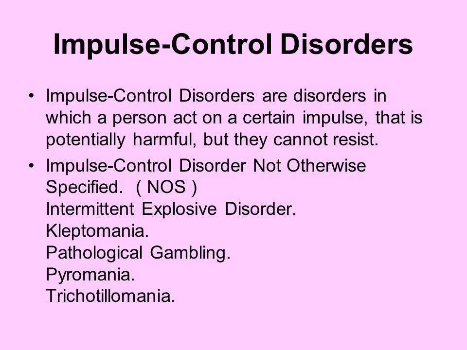
In 1978-1979. A.M. Markhashov under the direction of G.A. Ilizarov created a pin apparatus for fixing the vertebrae of dogs and began the development of an apparatus for fixing the human spine.

The studies were carried out from 1983 to 1985 and included many aspects. Researchers were: A.M. Markhashov, K.P. Kirsanov, Yu.A. Mushtaeva, G.D. Safonova, P.I. Kovalenko, L.O. Marchenkova, I.A. Menshchikov. The work opened a new page in spinal surgery.

External transpedicular fixation of the spine has become an innovative method for the treatment of patients with spinal pathology. Controlled dosed controlled extrafocal impact on the spine was based on the philosophy of the method of G.A. Ilizarov and was its continuation. This method of correcting spinal deformities of various etiologies with the possibility of creating conditions for spinal fusion was a pioneering alternative direction for existing fixation devices.

January 1993 a scientific group and a department of vertebrology and neurosurgery have been organized at the RRC VTO. The head of the department and the head of the clinical group was appointed Ph.D. Khudyaev Alexander Timofeevich. Under the leadership of A.T. Khudyaev, the use of an external transpedicular fixation apparatus was started in the treatment of patients with traumatic injuries of the spine, scoliotic and kyphotic deformities, and spondylolisthesis. In the years when modern submersible implants were not available in Russia, the external transpedicular fixation apparatus became the main tool in the treatment of patients with acute spinal cord and spinal cord injury, patients with the consequences of trauma, for the correction of scoliotic and kyphotic spinal deformities and spondylolisthesis.

In the future, the range of surgical interventions and applied methods of treating patients was constantly expanding. The main activities of the department were: the treatment of patients with spinal deformities of various etiologies, degenerative-dystrophic diseases and the use of neuromodulation techniques in patients with the consequences of damage to the spinal cord and peripheral nerves. Since January 2015, on the basis of the neurosurgical department, the traumatology and orthopedic department No. 10 has been formed.

Currently, the main clinical and scientific direction of the department's activity is the surgical treatment of adult patients with spinal deformities of various origins, degenerative diseases, spinal injuries, consequences of injuries and previous surgical interventions. For this, modern diagnostic tools, the latest treatment technologies and medical equipment are used.

The main concept of treatment

is not only surgical treatment of spinal pathology, but orthopedic, neurological and functional rehabilitation in general for patients with severe spinal pathology to improve the quality and life expectancy.

Employees

A decompressive and stabilizing intervention was performed from the anterior approach with removal of a C6-C7 disc herniation and disc prosthesis with an interbody cage.

Multilevel hernia and stenosis of the spinal canal of the cervical spine

Clinical example: Patient T., 52 years old, was admitted with complaints of

Diagnosis: Dorsopathy. Cervical osteochondrosis. Spinal stenosis at the level of C2 - C7. Vertebrogenic cervical radiculomyelopathy. Tetraparesis. NPF.

Completed: Microsurgical decompression of the dural sac with reconstruction of the spinal canal at the C3-C7 level. Spondylosynthesis C2-C7. Laminectomy of C3-C6 vertebrae, resection of the arch of C7 vertebrae. Posterior fixation of C2-Th2 vertebrae.

Lumbar disc herniation

Clinical example: Patient I., 34 years old

Diagnosis: Lumbar osteochondrosis. Spinal stenosis. Paramedian left-sided disc herniation L4-5. Radiculopathy S1 on the left.

Left L4-5 interlaminectomy performed. Microsurgical removal of disc herniation L4-5.

Stenosis of the spinal canal of the cervical spine

Clinical example: Patient T., 56 years old

Diagnosis: Atlanto-axial dislocation against the background of C1-C2 rheumatoid lesion. Stenosis of the spinal canal at the level of C1-C2. Vertebrogenic cervical myelopathy IIIA according to Ranawat. Spastic tetraparesis. Seropositive rheumatoid arthritis erosive stage Rg 4st.

Decompression and stabilization intervention performed. C1 laminectomy. Posterior fixation of C0-C2 vertebrae. Posterior local osteoplastic spondylodesis with autologous bone.

Spinal stenosis of the lumbar spine

Clinical example: Patient B., 51 years old

Diagnosis: Dorsopathy. Lumbar osteochondrosis. Degenerative antelisthesis of the L2 vertebra of the 1st degree, antelisthesis of the L3 vertebra of the 1st degree. Stenosis of the spinal canal at the level of L2 - L3 - L4 vertebrae. Neurogenic intermittent claudication. Vertebrogenic pain syndrome.

Completed Decompression and stabilization intervention. Reconstruction of the spinal canal at the level of L2, L3 vertebrae. PLIF cages segments L2 - L3, L3 - L4. Posterior fixation of L2, L3, L4 vertebrae with a transpedicular fixation system.

Lumbar stenosis

Clinical example: Patient T., 57 years old

Diagnosis: Dorsopathy. Combined spinal stenosis. Chronic lumbago. Neurogenic intermittent claudication.

Completed: reconstruction of the spinal canal due to distraction laminoplasty with implantation of a dynamic interspinous fixator.

- Spondylolisthesis of any etiology

Spondylolisthesis

Clinical example: Patient B., 51 years old

Diagnosis: Dorsopathy. Lumbar osteochondrosis. Degenerative unstable antelisthesis of the L2 vertebra of the 1st degree, antelisthesis of the L3 vertebra of the 1st degree. Stenosis of the spinal canal at the level of L2 - L3 - L4 vertebrae. Neurogenic intermittent claudication. Vertebrogenic pain syndrome.

Decompressive stabilization intervention performed. Reconstruction of the spinal canal at the level of L2, L3 vertebrae. PLIF caged segments L2 - L3, L3 - L4. Posterior fixation of L2, L3, L4 vertebrae with a transsedicular fixation system.

- Scoliosis in adults

Degenerative scoliosis

Clinical example: Patient D., 69 years old

Diagnosis: ASD. Dorsopathy. Lumbar osteochondrosis. Spinal stenosis at the level of L3-L5 vertebrae. Degenerative spondylolisthesis L4 vertebra 1 tbsp. Degenerative lumbar scoliosis type NBPM SRS-Schwab. Sagittal imbalance type 2. Vertebrogenic pain syndrome. Neurogenic intermittent claudication.

Decompressive and stabilizing surgery performed. Smith-Petersen osteotomy at the level of L2-L5 vertebrae with decompression of the dural sac at the level of L3-L5 vertebrae. Correction and posterior stabilization of the spine with a transpedicular fixation system.

Outcome of idiopathic scoliosis in adults

Clinical example: Patient O., 19 years old

Diagnosis: ASD. The outcome is scoliotic deformity of the thoracic, lumbar spine type 4C - according to Lenke. Right rib hump. Sagittal imbalance type 1. Vertebrogenic pain syndrome.

Performed a reconstructive intervention with multilevel vertebrotomy. Smith-Petersen osteotomy at Th5-8-9. Correction and posterior fixation of Th3-L3 with the Legasy and LegasyLiteMedtronic TPF system. The rear local bone-plastic spondylodisiness of

-kyphotic deformations of any etiology in adults

Kifotic spinal deformation against the backdrop of osteochondropathy Shoyerman-MAU 9000

Diagnosis: kyphotic deformation of the sistor on the phono ostechondrome. Sagittal imbalance type 1. Vertebrogenic pain syndrome.

Correction and stabilization of the spinal deformation by the tracsticular fixation system

- damage to the cervical, thoracic and lumbar spine in adults

Fractures of the craniwetrail zone

K. Diagnosis: Explosive fracture of the C1 vertebra of the Jefferson type. Atlanto - axial instability. Bulbar paresis. Vertebrogenic pain syndrome.

Decompressive-stabilizing surgery performed. Repositioning-stabilizing spondylosynthesis of the C1 vertebra.

Fractures of the craniovertebral zone

Clinical example: Patient O., aged 52

Diagnosis: An old fracture of the C2 tooth of the vertebra. Pseudarthrosis. Condition after surgical treatment. Pseudarthrosis. Vertebrogenic cervical myelopathy. Tetraparesis.

Decompressive and stabilizing intervention performed. Removal of wire fixation. Posterior fixation of C0-C3 vertebrae.

Injuries of the subaxial section

Clinical example: Patient S., 57 years old

Diagnosis: Traumatic disease of the spinal cord, acute period. Linked left-sided dislocation of the C3 vertebra. Traumatic disc herniation C3-C4. Easy tetraparesis. NPF.

Open reduction of dislocation of C3 vertebra was performed. Anterior spinal fusion C3-C4 PEEK cage with extraosseous plate.

Thoracic and lumbar spine injuries

Clinical example: Patient P., aged 18

Diagnosis: TBCI, acute period. Fracture-dislocation of Th22 vertebra, Tlc type C, N4, M1 (according to AO Spine). lower paraplegia. NPF.

A decompressive and stabilizing intervention was performed from the posterior approach. Open reduction of dislocation of Th22 vertebra, removal of traumatic disc herniation Th22-L1. Reconstruction of the spinal canal at the level of Th22-L1 vertebrae. Repositioning-stabilizing Th9-L2 spondylosynthesis with the Stryker TPF system. Anterior spinal fusion Th22-L1 with autobone. Posterior osteoplastic spondylodesis.

Posttraumatic deformities

Clinical example: Patient L., 35 years old

Diagnosis: TBCI, late period. Consequences of fractures of Th7, Th8 vertebrae, type C according to the AOSpine classification. Kyphotic deformity of the thoracic spine. Lower flaccid paraplegia. Dysfunction of the pelvic organs.

Decompressive-stabilizing surgery was performed. VCR-type vertebrotomy at the level of Th5, Th6 vertebrae. Correction and fixation of Th3-Th9vertebrae with a transpedicular fixation system and Th5-Th7 interbody fusion with a mesh implant.

Post-traumatic deformities

Clinical example: Patient K., 43 years old

Diagnosis: ASD. Severe kyphotic deformity of the thoracic and lumbar spine against the background of an old fracture of the L1 vertebra. Condition after surgical treatment. Sagittal imbalance type 2. Neurogenic intermittent claudication. Vertebrogenic pain syndrome.

A decompressive and stabilizing intervention was performed with resection of the vertebra from the posterior approach with spondylosynthesis of the spine. VCR-type vertebrotomy at the level of Th22-L1 vertebrae. Correction and posterior fixation of Th8-L4 with Xia2 Stryker TPF system. Interbody fusion with Mesh implant filled with autologous bone.


- Consequences of injuries of the spine and spinal cord in adults

Method of epidural electrical spinal cord stimulation (Patent No. 2441679 A61N 1/36 "Method of electrical stimulation of the spinal cord")

The treatment is aimed at stimulating the conduction pathways of the spinal cord, preserved after injury, in order to activate them and possibly improve motor and sensory functions.

Treatment includes an integrated approach with the involvement of a rehabilitation doctor, exercise therapy instructor, medical psychologist.

The method of electrical stimulation is aimed at activating the conduction of nerve impulses in damaged nerve endings, in connection with which the motor and sensory function of the limbs improves. The dynamics of positive changes depends on the nature of the spinal cord injury and is individual in each case. The method involves a minimally invasive (through a skin puncture) epidural installation of electrodes. In the future, electrostimulation sessions are carried out on the electrodes. After a course of stimulation (10-15 procedures), the electrodes are removed.

Indications for use:

  • Consequences of spinal cord injury with a syndrome of partial conduction disturbance with clinical manifestations of paresis of the limb muscles with (or) concomitant sensory, trophic disorders and dysfunction of the pelvic organs
  • Acute or intermediate TBCI
  • As a test (diagnostic) appointment in the late period of TBCI and in doubtful cases
  • Consequences of disorders of the spinal circulation
  • Pelvic organ dysfunction
  • Vascular myelopathy
  • Postoperative pain syndromes and neurological complications
  • Radiculopathy of various origins
  • Traumatic and postoperative injuries of peripheral nerves combined
  • Traction brachioplexopathies of various origins.

- Stenosis of the spinal canal on the background of diseases and injuries

Stenosis of the spinal canal of the cervical spine

Clinical example: Patient T., 56 years old

Diagnosis: Atlanto-axial dislocation on the background of C1-C2 rheumatoid lesion. Stenosis of the spinal canal at the level of C1-C2. Vertebrogenic cervical myelopathy IIIA according to Ranawat. Spastic tetraparesis. Seropositive rheumatoid arthritis erosive stage Rg 4st.

Decompression and stabilization intervention performed. C1 laminectomy. Posterior fixation of C0-C2 vertebrae. Posterior local osteoplastic spondylodesis with autologous bone.

Stenosis of the spinal lumbar spine

Clinical example: Patient B., 51 years old

Diagnosis: Dorsopathy. Lumbar osteochondrosis. Degenerative antelisthesis of the L2 vertebra of the 1st degree, antelisthesis of the L3 vertebra of the 1st degree. Stenosis of the spinal canal at the level of L2 - L3 - L4 vertebrae. Neurogenic intermittent claudication. Vertebrogenic pain syndrome.

Completed Decompression and stabilization intervention. Reconstruction of the spinal canal at the level of L2, L3 vertebrae. PLIF cages segments L2 - L3, L3 - L4. Posterior fixation of L2, L3, L4 vertebrae with a transpedicular fixation system.

Lumbar stenosis

Clinical example: Patient T., 57 years old

Diagnosis: Dorsopathy. Combined spinal stenosis. Chronic lumbago. Neurogenic intermittent claudication.

Completed: reconstruction of the spinal canal due to distraction laminoplasty with implantation of a dynamic interspinous fixator.

- Congenital malformations of the cervical spine and craniocervical region in adults;

- Pathology of the cervical spine in adults

Herniated cervical intervertebral discs

Clinical example: Patient K. , 39 years old

Diagnosis: Cervical osteochondrosis. Herniated disc C6-C7. Radicular syndrome C7 on the left.

A decompressive and stabilizing intervention was performed from the anterior approach with removal of a C6-C7 disc herniation and disc prosthesis with an interbody cage.

Multilevel hernia and stenosis of the spinal canal of the cervical spine

Clinical example: Patient T., 52 years old, was admitted with complaints of

Diagnosis: Dorsopathy. Cervical osteochondrosis. Spinal stenosis at the level of C2 - C7. Vertebrogenic cervical radiculomyelopathy. Tetraparesis. NPF.

Completed: Microsurgical decompression of the dural sac with reconstruction of the spinal canal at the C3-C7 level. Spondylosynthesis C2-C7. Laminectomy of C3-C6 vertebrae, resection of the arch of C7 vertebrae. Posterior fixation of C2-Th2 vertebrae.

Stenosis of the spinal canal of the cervical spine

Clinical example: Patient T. , 56 years old

Diagnosis: Atlanto-axial dislocation against the background of C1-C2 rheumatoid lesion. Stenosis of the spinal canal at the level of C1-C2. Vertebrogenic cervical myelopathy IIIA according to Ranawat. Spastic tetraparesis. Seropositive rheumatoid arthritis erosive stage Rg 4st.

Decompression and stabilization intervention performed. C1 laminectomy. Posterior fixation of C0-C2 vertebrae. Posterior local osteoplastic spondylodesis with autologous bone.

Injuries of the cervical spine

Fractures of the craniovertebral zone

Clinical example: Patient K., aged 47

Diagnosis: Explosive fracture of C1 type Jefferson vertebra. Atlanto - axial instability. Bulbar paresis. Vertebrogenic pain syndrome.

Decompressive-stabilizing surgery performed. Repositioning-stabilizing spondylosynthesis of the C1 vertebra.

Fractures of the craniovertebral zone

Clinical example: Patient O. , aged 52

Diagnosis: An old fracture of the C2 tooth of the vertebra. Pseudarthrosis. Condition after surgical treatment. Pseudarthrosis. Vertebrogenic cervical myelopathy. Tetraparesis.

Decompressive and stabilizing intervention performed. Removal of wire fixation. Posterior fixation of C0-C3 vertebrae.

Injuries of the subaxial section

Clinical example: Patient S., 57 years old

Diagnosis: Traumatic disease of the spinal cord, acute period. Linked left-sided dislocation of the C3 vertebra. Traumatic disc herniation C3-C4. Easy tetraparesis. NPF.

Open reduction of dislocation of C3 vertebra was performed. Anterior spinal fusion C3-C4 PEEK cage with extraosseous plate.

- Congenital deformities of the spine in adults;

- Tumors of the spine and spinal cord in adults

Tumors of the spinal cord. Clinical example: Patient M. , aged 51

Diagnosis: Intradural extramedullary neoplasm of the spinal canal at the level of Th5, Th6. Thoracicalgia.

Intervention performed: Laminectomy of the Th5 vertebrae, partial resection of the Th6 vertebral arch plate. Removal of an intradural extramedullary volumetric formation at the level of Th5, Th6 using microsurgical techniques. Stabilization of the spine with a transpedicular fixation system.

Spinal tumors. Clinical example: Patient M, 54 years old

Diagnosis: Hemangiomas of Th21 bodies, 12 vertebrae

Completed: Transcutaneous puncture monoportal vertebroplasty of Th21 vertebra on the right with bone cement. Transcutaneous puncture biportal vertebroplasty of the L2 vertebra with bone cement.

- Conditions after surgical interventions: failure of spinal fixation systems, fractures of structural elements, continued deformities, progression of deformities

Post-traumatic deformities
Clinical example: Patient K. , aged 43
Diagnosis: ASD. Severe kyphotic deformity of the thoracic and lumbar spine against the background of an old fracture of the L1 vertebra. Condition after surgical treatment. Sagittal imbalance type 2. Neurogenic intermittent claudication. Vertebrogenic pain syndrome.
A decompressive-stabilizing intervention was performed with resection of the vertebra from the posterior approach with spondylosynthesis of the spine. VCR-type vertebrotomy at the level of Th22-L1 vertebrae. Correction and posterior fixation of Th8-L4 with Xia2 Stryker TPF system. Interbody fusion with a Mesh implant filled with autologous bone.

Revision interventions: failure of spinal fixation systems
Clinical example: Patient S., 44 years old
Diagnosis: Consequences of a fracture of the L2 vertebra. Condition after surgical treatment. Failure of the metal structure. Chronic lumbago.
A decompressive-stabilizing intervention was performed with resection of L2, L3 vertebrae according to the VCR type. Dismantling of the TPF system, removal of a fragment of the screw L3 of the vertebra on the left. Repositioning-stabilizing Th22-L3 spondylosynthesis with a mesh endoprosthesis, Th21-L4 spondylosynthesis with a transpedicular fixation system.

Clinical example: Patient B., aged 29
Diagnosis: TBCM, late period. Sequelae of fracture-dislocation of the Th22 vertebra. Condition after surgical treatment. Instability of the metal structure. Lower gross paraparesis.
Reconstruction of the spinal canal at the level of Th22-L1 segments was performed. Dismantling of the TPF system, removal of fragments of screws of the L2 vertebra. Curettage of the disc Th22-L1. TLIF Th22-L1 cage. Correction and posterior fixation of Th21-L2 with a transpedicular fixation system.

- Spinal deformities due to Parkinson's disease

Clinical example: Patient I., 52 years old

Diagnosis: ASD. Kyphoscoliotic deformity of the thoracic and lumbar spine due to Parkinson's disease . Leaning Tower of Pisa Syndrome. Sagittal imbalance type 2. Frontal imbalance type 2.

Surgical treatment performed: Posterior fixation of Th5-S1 vertebrae and pelvic bones with a transpedicular fixation system. Posterior local osteoplastic fusion

- Deformities of the spine due to ankylosing spondylitis (Bekhterev's disease)

Clinical example: Patient K., 52 years old

Diagnosis: Ankylosing spondylitis. Kyphotic deformity of the spine, type IIA+. Combined stenosis of the spinal canal. Left-sided mild mixed hemiparesis.

Surgical treatment performed: Correction and posterior fixation of the spine with a transpedicular fixation system at the level of Th9--L5 vertebrae. Smith-Petersen osteotomy at the level of Th20-11, Th21-12, Th22-L1, L1-L2, L2-L3 vertebrae. Posterior local osteoplastic fusion.


- Spinal deformities due to rheumatoid arthritis

Spinal deformities due to rheumatoid arthritis

Clinical example: Patient B. , aged 37

Diagnosis: ASD. Severe kyphoscoliotic deformity of the cervical, thoracic and lumbar spine. Condition after surgical treatment. Combined limb deformities. Vertebrogenic cervical myelopathy. Tetraparesis. NPF.

Rheumatoid arthritis, seronegative, articular form, ACCP negative, late stage, activity 2 stage 4. FC IV.

Systemic osteoporosis. ICD. Chronic pyelonephritis without exacerbation.

Two stages of treatment were performed: Stage 1: application of a halo crown followed by traction.

Stage 2: reconstructive intervention with repositioning and stabilizing spondylosynthesis of SHOP and GOP. Correction and posterior fixation of C2-Th6 vertebrae using fixation systems for the cervical and thoracic spine.


- Consequences of damage to the peripheral nervous system

Department closely cooperates with:

  • X-ray department
  • Department of Anesthesiology and Intensive Care
  • Department of Traumatology and Orthopedics No. 9 (Department of Pathology of the Axial Skeleton and Neurovertebrology of Children),
  • Scientific laboratory of the Clinic for Spinal Pathology and Rare Diseases

The department has 36 beds. There are three superior rooms. Treatment is carried out under the programs of VMP, CHI, VHI and on a paid basis

Modern X-ray, neurophysiological, functional equipment and MRI are used for diagnostics.

The tactics of treating each patient is determined individually based on the examination, taking into account comorbidities and on the basis of world standards and approaches to surgical and rehabilitation treatment.

Operating rooms are equipped with modern medical equipment and spinal fixation systems. To control the state of the spinal cord during the operation, a mandatory neurophysiological control is carried out.

In the postoperative period, infusion drug therapy is carried out and the process of rehabilitation of patients takes place under the guidance and with the help of physical therapy instructors.

Prudnikova Oksana Germanovna

Head of the traumatological and orthopedic department No. 10. Doctor of Medical Sciences, neurosurgeon of the highest category, orthopedist-traumatologist, vertebrologist. Leading Researcher, Scientific Clinical Experimental Laboratory of Axial Skeleton Pathology and Neurosurgery

Summary Download

https://www.researchgate.net/profile/Oxana_Prudnikova?ev=prf_highl

Contacts: (3522) 45 33 57, [email protected]

Evsyukov Alexey Vladimirovich

Candidate of Medical Sciences, neurosurgeon.

Head of the Clinic for Spinal Pathology and Rare Diseases of the Ilizarov Center

Khomchenkov Maxim Viktorovich

Orthopedist-traumatologist, vertebrologist

Junior Researcher, Scientific Clinical Experimental Laboratory of Axial Skeleton Pathology and Neurosurgery

Resume download

Kotelnikov Alexander Olegovich

Traumatologist-orthopedist

Garipov Ilgiz Ildarovich

Orthopedist-traumatologist, PhD student

Volynsky Alexey Leonidovich

Neurosurgeon

Kuzmenko Elmira Ismailovna

Senior Nurse, Highest qualification category.

Work experience at the Center since 2011.

Diploma in the specialty "Medicine", qualification of a paramedic 1996.

Khlystova Yulia Alexandrovna

Specialist LLP No. 10

Operating since 2006

Education: Higher pedagogical education


Learn more