Facts about neurons in the brain
The Neuron
- Published1 Apr 2012
- Reviewed1 Apr 2012
- Author
- Source BrainFacts/SfN
Cells within the nervous system, called neurons, communicate with each other in unique ways. The neuron is the basic working unit of the brain, a specialized cell designed to transmit information to other nerve cells, muscle, or gland cells.
Neurons are cells within the nervous system that transmit information to other nerve cells, muscle, or gland cells. Most neurons have a cell body, an axon, and dendrites. The cell body contains the nucleus and cytoplasm. The axon extends from the cell body and often gives rise to many smaller branches before ending at nerve terminals.
Dendrites extend from the neuron cell body and receive messages from other neurons. Synapses are the contact points where one neuron communicates with another. The dendrites are covered with synapses formed by the ends of axons from other neurons.
Illustration by Lydia V. Kibiuk, Baltimore, MD; Devon Stuart, Harrisburg, PA
Neurons are cells within the nervous system that transmit information to other nerve cells, muscle, or gland cells. Most neurons have a cell body, an axon, and dendrites. The cell body contains the nucleus and cytoplasm. The axon extends from the cell body and often gives rise to many smaller branches before ending at nerve terminals. Dendrites extend from the neuron cell body and receive messages from other neurons. Synapses are the contact points where one neuron communicates with another. The dendrites are covered with synapses formed by the ends of axons from other neurons.
The brain is what it is because of the structural and functional properties of interconnected neurons.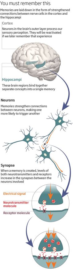 The mammalian brain contains between 100 million and 100 billion neurons, depending on the species. Each mammalian neuron consists of a cell body, dendrites, and an axon.
The cell body contains the nucleus and cytoplasm. The axon extends from the cell body and often gives rise to many smaller branches before ending at nerve terminals.
Dendrites extend from the neuron cell body and receive messages from other neurons. Synapses are the contact points where one neuron communicates with another. The dendrites are covered with synapses formed by the ends of axons from other neurons.
When neurons receive or send messages, they transmit electrical impulses along their axons, which can range in length from a tiny fraction of an inch (or centimeter) to three feet (about one meter) or more. Many axons are covered with a layered myelin sheath, which accelerates the transmission of electrical signals along the axon. This sheath is made by specialized cells called glia. In the brain, the glia that make the sheath are called oligodendrocytes, and in the peripheral nervous system, they are known as Schwann cells.
The brain contains at least ten times more glia than neurons. Glia perform many jobs. Researchers have known for a while that glia transport nutrients to neurons, clean up brain debris, digest parts of dead neurons, and help hold neurons in place. Current research is uncovering important new roles for glia in brain function.
Content Provided By
Related Topics Neurons Cells & Circuits Brain Anatomy & Function
Brain Basics: The Life and Death of a Neuron
Image
Until recently, most neuroscientists thought we were born with all the neurons we were ever going to have. As children we might produce some new neurons to help build the pathways - called neural circuits - that act as information highways between different areas of the brain. But scientists believed that once a neural circuit was in place, adding any new neurons would disrupt the flow of information and disable the brain’s communication system.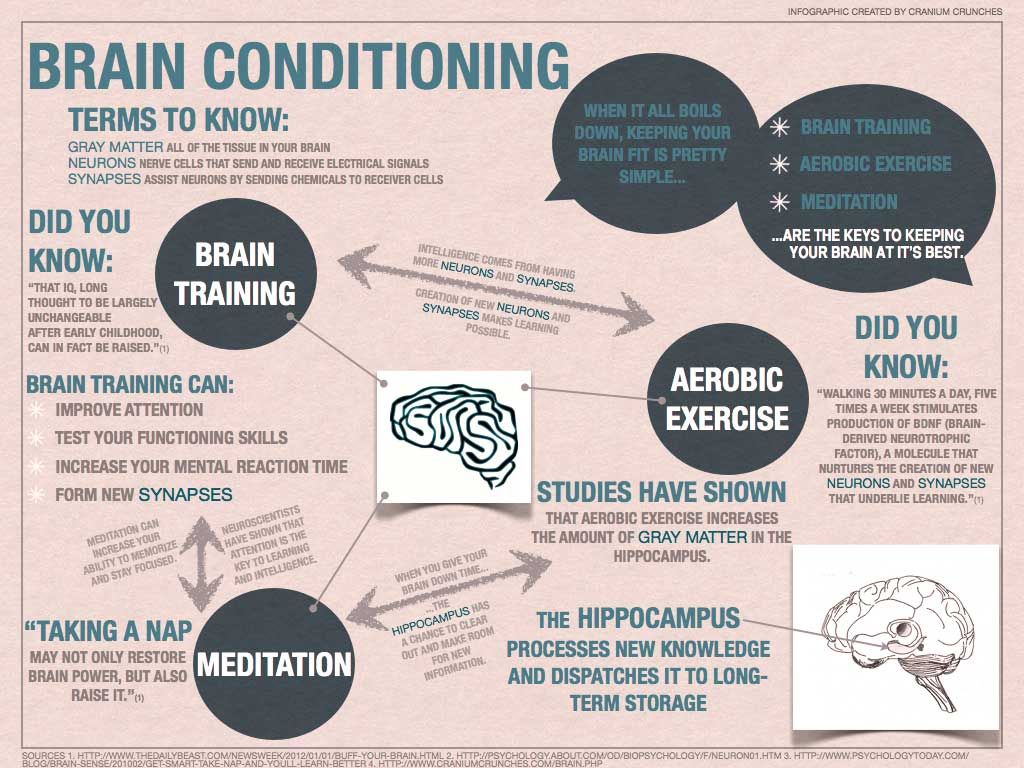
In 1962, scientist Joseph Altman challenged this belief when he saw evidence of neurogenesis (the birth of neurons) in a region of the adult rat brain called the hippocampus. He later reported that newborn neurons migrated from their birthplace in the hippocampus to other parts of the brain. In 1979, another scientist, Michael Kaplan, confirmed Altman’s findings in the rat brain, and in 1983 he found neural precursor cells in the forebrain of an adult monkey.
These discoveries about neurogenesis in the adult brain were surprising to other researchers who didn’t think they could be true in humans. But in the early 1980s, a scientist trying to understand how birds learn to sing suggested that neuroscientists look again at neurogenesis in the adult brain and begin to see how it might make sense. In a series of experiments, Fernando Nottebohm and his research team showed that the numbers of neurons in the forebrains of male canaries dramatically increased during the mating season.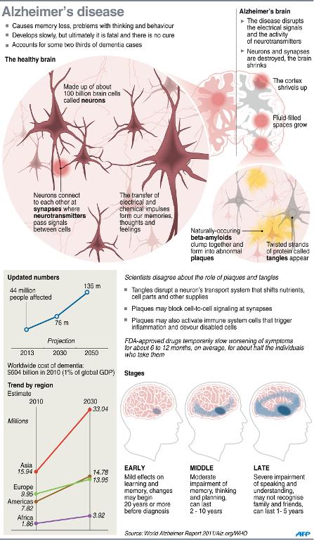 This was the same time in which the birds had to learn new songs to attract females.
Why did these bird brains add neurons at such a critical time in learning? Nottebohm believed it was because fresh neurons helped store new song patterns within the neural circuits of the forebrain, the area of the brain that controls complex behaviors. These new neurons made learning possible. If birds made new neurons to help them remember and learn, Nottebohm thought the brains of mammals might too.
Other scientists believed these findings could not apply to mammals, but Elizabeth Gould later found evidence of newborn neurons in a distinct area of the brain in monkeys, and Fred Gage and Peter Eriksson showed that the adult human brain produced new neurons in a similar area.
For some neuroscientists, neurogenesis in the adult brain is still an unproven theory. But others think the evidence offers intriguing possibilities about the role of adult-generated neurons in learning and memory.
The Architecture of the Neuron
The central nervous system (which includes the brain and spinal cord) is made up of two basic types of cells: neurons (1) and glia (4) & (6). Glia outnumber neurons in some parts of the brain, but neurons are the key players in the brain.
Neurons are information messengers. They use electrical impulses and chemical signals to transmit information between different areas of the brain, and between the brain and the rest of the nervous system. Everything we think and feel and do would be impossible without the work of neurons and their support cells, the glial cells called astrocytes (4) and oligodendrocytes (6).
Neurons have three basic parts: a cell body and two extensions called an axon (5) and a dendrite (3). Within the cell body is a nucleus (2), which controls the cell’s activities and contains the cell’s genetic material. The axon looks like a long tail and transmits messages from the cell. Dendrites look like the branches of a tree and receive messages for the cell. Neurons communicate with each other by sending chemicals, called neurotransmitters, across a tiny space, called a synapse, between the axons and dendrites of adjacent neurons.
Image
There are three classes of neurons:
- Sensory neurons carry information from the sense organs (such as the eyes and ears) to the brain.
- Motor neurons control voluntary muscle activity such as speaking and carry messages from nerve cells in the brain to the muscles.
- All the other neurons are called interneurons.
Scientists think that neurons are the most diverse kind of cell in the body. Within these three classes of neurons are hundreds of different types, each with specific message-carrying abilities.
How these neurons communicate with each other by making connections is what makes each of us unique in how we think, and feel, and act.
Birth
The extent to which new neurons are generated in the brain is a controversial subject among neuroscientists. Although the majority of neurons are already present in our brains by the time we are born, there is evidence to support that neurogenesis (the scientific word for the birth of neurons) is a lifelong process.
Neurons are born in areas of the brain that are rich in concentrations of neural precursor cells (also called neural stem cells). These cells have the potential to generate most, if not all, of the different types of neurons and glia found in the brain.
Neuroscientists have observed how neural precursor cells behave in the laboratory. Although this may not be exactly how these cells behave when they are in the brain, it gives us information about how they could be behaving when they are in the brain’s environment.
The science of stem cells is still very new, and could change with additional discoveries, but researchers have learned enough to be able to describe how neural stem cells generate the other cells of the brain. They call it a stem cell’s lineage and it is similar in principle to a family tree.
Neural stem cells increase by dividing in two and producing either two new stem cells, or two early progenitor cells, or one of each.
When a stem cell divides to produce another stem cell, it is said to self-renew. This new cell has the potential to make more stem cells.
When a stem cell divides to produce an early progenitor cell, it is said to differentiate. Differentiation means that the new cell is more specialized in form and function. An early progenitor cell does not have the potential of a stem cell to make many different types of cells. It can only make cells in its particular lineage.
Early progenitor cells can self-renew or go in either of two ways. One type will give rise to astrocytes. The other type will ultimately produce neurons or oligodendrocytes.
Migration
Once a neuron is born it has to travel to the place in the brain where it will do its work.
How does a neuron know where to go? What helps it get there?
Scientists have seen that neurons use at least two different methods to travel:
- Some neurons migrate by following the long fibers of cells called radial glia.
 These fibers extend from the inner layers to the outer layers of the brain. Neurons glide along the fibers until they reach their destination.
 - Neurons also travel by using chemical signals. Scientists have found special molecules on the surface of neurons -- adhesion molecules -- that bind with similar molecules on nearby glial cells or nerve axons. These chemical signals guide the neuron to its final location.
Not all neurons are successful in their journey. Scientists think that only a third reach their destination. Some cells die during the process of neuronal development.
Some neurons survive the trip, but end up where they shouldn’t be. Mutations in the genes that control migration create areas of misplaced or oddly formed neurons that can cause disorders such as childhood epilepsy. Some researchers suspect that schizophrenia and the learning disorder dyslexia are partly the result of misguided neurons.
Image
Differentiation
Image
Once a neuron reaches its destination, it has to settle in to work. This final step of differentiation is the least well-understood part of neurogenesis.
Neurons are responsible for the transport and uptake of neurotransmitters - chemicals that relay information between brain cells.
Depending on its location, a neuron can perform the job of a sensory neuron, a motor neuron, or an interneuron, sending and receiving specific neurotransmitters.
In the developing brain, a neuron depends on molecular signals from other cells, such as astrocytes, to determine its shape and location, the kind of transmitter it produces, and to which other neurons it will connect. These freshly born cells establish neural circuits - or information pathways connecting neuron to neuron - that will be in place throughout adulthood.
But in the adult brain, neural circuits are already developed and neurons must find a way to fit in. As a new neuron settles in, it starts to look like surrounding cells. It develops an axon and dendrites and begins to communicate with its neighbors.
Death
Although neurons are the longest living cells in the body, large numbers of them die during migration and differentiation.
The lives of some neurons can take abnormal turns. Some diseases of the brain are the result of the unnatural deaths of neurons.
- In Parkinson’s disease, neurons that produce the neurotransmitter dopamine die off in the basal ganglia, an area of the brain that controls body movements. This causes difficulty initiating movement.
- In Huntington’s disease, a genetic mutation causes over-production of a neurotransmitter called glutamate, which kills neurons in the basal ganglia. As a result, people twist and writhe uncontrollably.
- In Alzheimer’s disease, unusual proteins build up in and around neurons in the neocortex and hippocampus, parts of the brain that control memory. When these neurons die, people lose their capacity to remember and their ability to do everyday tasks.
 Physical damage to the brain and other parts of the central nervous system can also kill or disable neurons.
 - Blows to the brain, or the damage caused by a stroke, can kill neurons outright or slowly starve them of the oxygen and nutrients they need to survive.
- Spinal cord injury can disrupt communication between the brain and muscles when neurons lose their connection to axons located below the site of injury. These neurons may still live, but they lose their ability to communicate.
Image
A diseased neuronImage
A dying neuron
Hope Through Research
Scientists hope that by understanding more about the life and death of neurons they can develop new treatments, and possibly even cures, for brain diseases and disorders that affect the lives of millions of Americans.
The most current research suggests that neural stem cells can generate many, if not all, of the different types of neurons found in the brain and the nervous system. Learning how to manipulate these stem cells in the laboratory into specific types of neurons could produce a fresh supply of brain cells to replace those that have died or been damaged.
Therapies could also be created to take advantage of growth factors and other signaling mechanisms inside the brain that tell precursor cells to make new neurons. This would make it possible to repair, reshape, and renew the brain from within.
10 facts about neurons
The expression "Nerve cells do not regenerate" was perceived as an indisputable truth quite recently. However, as it turned out, this is not entirely true: scientists have already found where the stocks of new neurons lie (in the hippocampus and olfactory bulb), and are thinking with might and main about how to further stimulate their recovery.
Science is moving forward, and it looks like it's time to brush up on the knowledge of neurons - the most excitable cells in the human body that process, store and transmit gigabytes of information throughout our body every second. nine0005
We have collected 10 interesting and informative facts for you: scroll through the gallery and find out even more
Despite the fact that the absolute irrecoverability of nerve cells is already in question, there is no doubt about the other: we lose them regularly. Moreover, "nervous stress" is experienced not only by adults loaded with work, but even by infants. At the same time, in the process of growing up, children lose significantly more neurons than their parents.
Neurons come in many shapes and sizes, depending on where they are in the body and what they are programmed for. Thus, neurons consist of a central cell and neurite processes, which can be located in very different ways. nine0005
All neurons are programmed to do different things. Sensory deliver electrical signals from external parts of the body to the central nervous system, and motor - vice versa. Receptor neurons perceive the environment (light, sound, etc.) and transmit data to sensory neurons, while interneurons send messages from one neuron to another.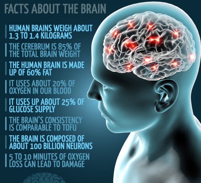
Neurons not only mobilize us to accomplish things: the parasympathetic nervous system also controls bodily functions when a person is at rest. For example, it includes stimulating digestion, activating metabolism, and helping with relaxation. nine0005
Scientists are developing ways to hack the immune system to control brain cells with flashes of light. The hack could help scientists learn more about the functions of different groups of neurons, enabling them to activate multiple brain cells at the same time and observe their effects on the body.
Neural connections are formed throughout life. Thanks to the beaten path of neural connections, we easily and automatically perform habitual actions. Breaking ties and abandoning old habits is not easy, however, with regular training, we force the brain to form new habits! nine0005
In the case of neurons, only young nerve cells, neuroblasts, can divide. When neurons reach maturity, they forget about this possibility and do not reproduce themselves by dividing, as other cells in our body do.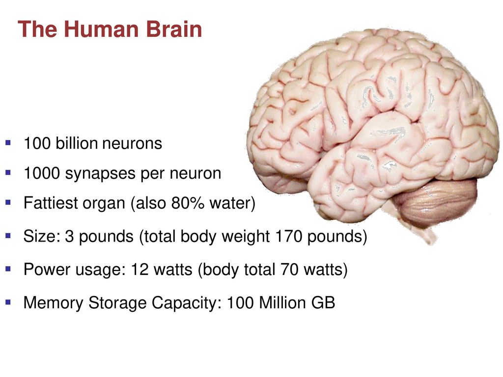
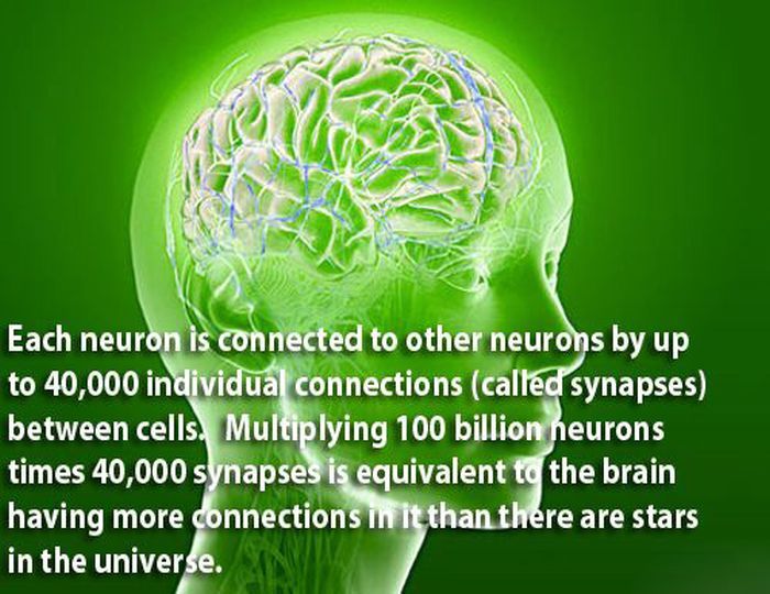
Our brain contains about 100 billion neurons, more than the number of stars in the Milky Way. And yet this is only 10% of all brain cells that we have.
Each neuron in our brain has somewhere between 1,000 and 10,000 connections to other neurons in the brain, bringing the total number of neural connections in the brain to 100 trillion. nine0005
Our memory, basic reflexes, abstract thoughts and even emotions are all products of the neural network in our body. Neurons are very special and require more glucose and oxygen than the rest of the cells in our body.
Anna Verelko
Tags
#Cards
#FACTS
#Science
#Brain
#body
#nerve cells
#nervous system
9000Five Scientific facts about the brain - News - News – IQ Research and Education Portal – National Research University Higher School of Economics
Neurons drive the crowd
The “joining the majority” effect, known to political technologists, can be explained by a feature of the brain that allowed a person to survive in the process of evolution.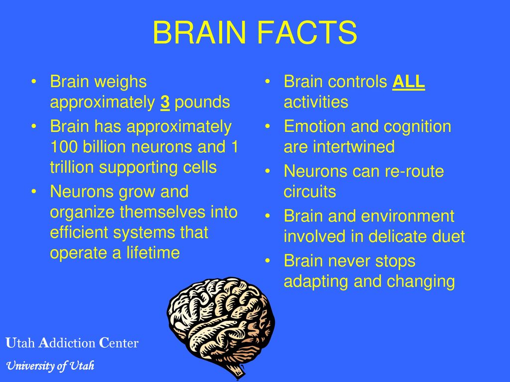 When a person is different from others, he receives an automatic signal of a “mistake”, which prompts him to change his opinion towards the majority. The fact that a “dopamine storm” occurs in the head at the moment of a change of opinion under the influence of others has been proven in the laboratory. nine0005
The choice of reward is related to the features of the brain
A person often acts impulsively when making a decision regarding the time of reward. Preference is given to instant reward, even if delayed in time looks more profitable. This can be explained from the point of view of the features of the brain, namely the degree of activity of its center responsible for reward - the ventral striatum. Therefore, according to neuroeconomic research, one person may be more patient in anticipation of a reward, while another is not. nine0005
Men are slower to switch from one task to another
When it becomes necessary to switch attention to another task, the brain of men begins to consume more energy than the brain of women. There is stronger activation in the dorsolateral prefrontal areas (that is, in the frontal cortex of the cerebral hemispheres, which is associated with areas that provide attention, cognitive activity and motor skills), as well as in those in which women do not activate. Such differences are typical for young men and women aged 20 to 45 years. nine0005
Learning foreign languages increases brain plasticity
The more foreign languages a person knows, the better he learns the data received in the learning process. Its neural networks that encode information about new words form faster. This is a promising line of research. Scientists believe that understanding the mechanisms of the functioning of the brain when learning languages will help to more effectively deal with speech disorders resulting from injuries or strokes. nine0005
"Owls" work more efficiently at night
"Larks" in the moonlight faster than "owls" cope with non-standard tasks for attention.