Parts of the brain amygdala
Limbic System: Amygdala (Section 4, Chapter 6) Neuroscience Online: An Electronic Textbook for the Neurosciences | Department of Neurobiology and Anatomy
6.1 Amygdala - General Considerations
Amygdala is the integrative center for emotions, emotional behavior, and motivation. If the brain is turned upside down the end of the structure continuous with the hippocampus is called the uncus. If you peel away uncus you will expose the amygdala which abuts the anterior of the hippocampus. Just like with the hippocampus, major pathways communicate bidirectionally and contain both efferent and afferent fibers.
Figure 6.1 |
6.2 Inputs to the Amygdala
Figure 6.2 |
As was the case with the hippocampus, fibers carrying inputs to the amygdala are in virtually all cases combined with fibers carrying outputs from the amygdala.
The amygdala receives inputs from all senses as well as visceral inputs. Since the amygdala is very important in emotional learning it is not surprising that visceral inputs are a major input source. Visceral inputs come from the hypothalamus, septal area, orbital cortex, and parabrachial nucleus. Olfactory sensory information comes from the olfactory bulb. Auditory, visual and somatosensory information comes from the temporal and anterior cingulate cortices.
Figure 6.3 |
6.3 Major Output Pathways of the Amygdala
- Ventral amygdalofugal pathway
- Stria terminalis
- Directly to the hippocampus
- Directly to the entorhinal cortex
- Directly to the dorsomedial nucleus of the thalamus
6. 4 Ventral Amygdalofugal Pathway
Ventral Amygdalofugal Pathway. The term "fugal" comes from the word fuge—to drive away—as in fugitive. This pathway continues to the anterior olfactory nucleus, anterior perforated substance, piriform cortex, orbitofrontal cortex, anterior cingulate cortex, and ventral striatum. The ventral striatum includes part of the caudate, putamen, and the nucleus accumbens septi (nucleus that reclines on the septum). Projections from the ventral striatum are links in a basal ganglia circuit that are important in stimulus-response associative learning. The ventral amygdalofugal pathway also connects to the hypothalamus and septal nucleus, but the amygdala's major connection to the hypothalamus and septal nucleus is through the stria terminalis.
The ventral amygdalofugal pathway is important because it is a link whereby motivation and drives, through the limbic system, can influence responses. It is also a link whereby responses are learned. In this case this is the link whereby associative learning takes place. That is where responses are associated with appetitive and aversive consequences that is rewards and punishers.
Three simplifications:
- The stria terminalis is similar in form, function, and location as the fornix for the hippocampal pathway. Thus by way of analogy one can say that the stria terminalis is to the amygdala as the fornix is to the hippocampus. Stria is a Latin word that means line, groove, or band. Related to the word "striated".
- The stria terminalis connects only to subcortical structures. (Connection to cortical structures is through the ventral amygdalofugal pathway.)
- The stria terminalis overlaps with the ventral amygdalofugal pathway in that it also connects to the septal nuclei and hypothalamus and thus forms a loop.
More on similarities to the fornix:
Like the fornix, the stria terminalis has precommissural and postcommissural branches in relation to the anterior commissure. The precommissural branch goes to the septal area. This is exactly what the fornix does. The postcommissural branch goes to the hypothalamus. This is exactly what the fornix does. Whereas the postcommissural branch of the fornix projects to mammillary bodies of the hypothalamus, the postcommissural branch of the stria terminalis projects to the lateral nucleus and ventral-medial nucleus of the hypothalamus.
As with the fornix, some fibers enter anterior commissure cross to the contralateral side. Just as in the case of the two hippocampi communicating with each other through the anterior commissure, the two amygdala communicate with each other through the anterior commissure.
The stria terminalis also projects to the habenula, which is part of the epithalamus.
The central nucleus of the amygdala produces autonomic components of emotion (e.g., changes in heart rate, blood pressure, and respiration) primarily through output pathways to the lateral hypothalamus and brain stem.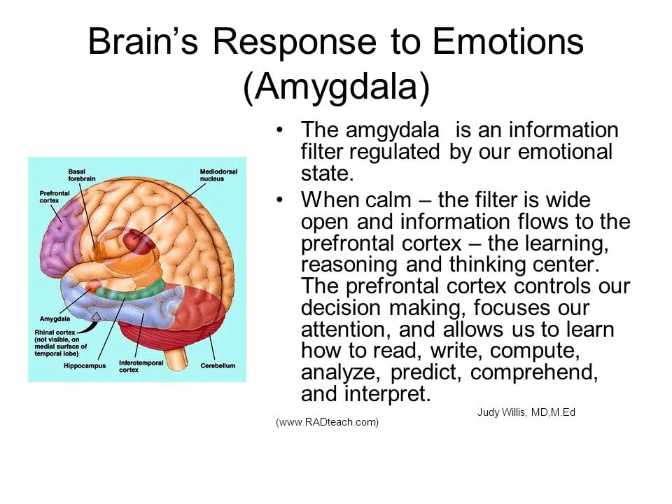
The central nucleus of the amygdala also produces conscious perception of emotion primarily through the ventral amygdalofugal output pathway to the anterior cingulate cortex, orbitofrontal cortex, and prefrontal cortex.
6.5 More on Function of the Amygdala
Stimulation of the amygdala causes intense emotion, such as aggression or fear.
Irritative lesions of temporal lobe epilepsy have the effect of stimulating the amygdala. In its extreme form irritative lesions of temporal lobe epilepsy can cause a panic attack. Panic attacks are brief spontaneously recurrent episodes of terror that generate a sense of impending disaster without a clearly identifiable cause. PET scans have shown an increase in blood flow to the parahippocampal gyri, beginning with the right parahippocampal gyrus. Similar but attenuated blood flow increases occurs during anxiety attacks.
Destructive lesions such as ablation of the amygdala cause an effect opposite to the irritative lesions of temporal lobe epilepsy. Destructive lesions of the amygdala cause tameness in animals, and a placid calmness in humans characterized as a flatness of affect. Lesions of the amygdala can occur as a result of Urbach-Wiethe disease where calcium is deposited in the amygdala. If this disease occurs early in life then these patients with bilateral amygdala lesions cannot discriminate emotion in facial expressions, but their ability to identify faces remains. The anatomical area for face recognition and memory is in the multimodal association area of the inferotemporal cortex. This is a good example of how emotion in one area (amygdala) is linked with perception in another area (inferotemporal cortex) to create an intense emotionally charged memory.
Figure 6.4 |
Flatness of affect is one of the symptoms of the previously mentioned Kluver-Bucy syndrome where the entire temporal lobes of monkeys were removed. Actually,just lesions of the amygdala were shown to be primarily responsible for flatness of affect. This work eventually led to the psychosurgical technique of prefrontal lobotomies. Remember the movie with Jack Nicholson, “One Flew Over the Cuckoo’s Nest.” The prefrontal cortex inputs into the amygdala. By severing this input a flatness of affect is produced which was thought to be desirable in schizophrenic patients who were aggressively violent or emotionally agitated.
The amygdala combines many different sensory inputs. Like the hippocampus it combines external and internal stimuli. Every sensory modality has input. These are integrated with somatosensory and visceral inputs—this is where you get your “gut reaction”. The link between prefrontal cortex, septal area, hypothalamus, and amygdala likely gives us our gut feelings, those subjective feelings, about what is good and what is bad.
It is also where memory and emotions are combined. When the reward is particularly sweet, that behavior and association may last a lifetime. Likewise, the trauma and humiliation of punishment may be remembered for a long time too.
6.6 Fear Conditioning: An Example of the Role of the Amygdala in Learning
Another example of emotion being linked to some perceptual experience is fear conditioning. In this example the sensory experience is auditory rather than visual as in the emotion of faces. Much of what we know about the amygdala and its role in emotional learning and memory comes from fear conditioning, mostly but not exclusively conducted with animals. This is an example of classical conditioning or Pavlovian conditioning. In the classic experiments conducted by Pavlov just after the turn of the century, a neutral stimulus—a bell—was sounded and after a brief interval food powder—the unconditioned stimulus—was placed in the dog’s mouth. After a few such pairings the dog would salivate to the sound of the bell. The crucial aspect of classical conditioning is that it is a pairing between two stimuli. No response is required to get the reward. In fear conditioning, an organism hears a noise or sees a visual stimulus. A few seconds, later it receives a mild shock. The reactions involve freezing, elevated blood pressure and heart rate, and it gets twitchy—startles easily.
Figure 6.5
|
||
Figure 6.6 (top) and 6.7 (bottom) |
Pathways from the thalamus to the amygdala are particularly important in emotional learning. Output pathways from the central nucleus of the amygdala make extensive connections with the brain stem for emotional responses and extensive connections with cortical areas through the nucleus basalis. Cholinergic projections from the nucleus basalis to the cortex are thought to arouse the cortex.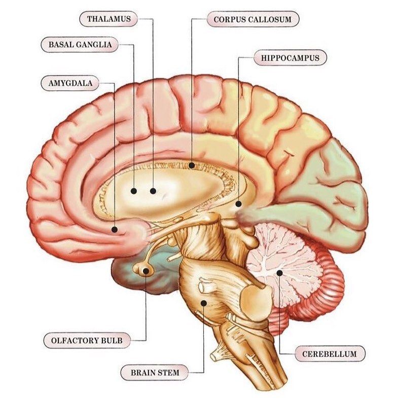
The following diagram provides additional information on outputs controlled by the amygdala during fear conditioning.
Figure 6.8 |
Some pathways of fear conditioning have been discovered and this is a hot research topic in neuroscience. If the auditory cortex pathway is lesioned, for example, basic fear conditioning is unaltered, but discrimination is altered. In the discrimination procedure one sound is paired with shock and another sound is not paired with shock. The animals had to rely solely on the thalamus and amygdala for learning and they could not learn the discrimination; apparently the two stimuli were indistinguishable.
So, the cortex is not needed for simple fear conditioning; instead it allows us to recognize an object by sight or sound— to interpret the environment.
Thus, pathways from the sensory thalamus provide only a crude perception of the world, but because they involve only one neural link they are fast pathways. Why might FAST be important? We need a quick reaction to potential danger. The thalamus—amygdala pathway provides us with this and may also prepare the amygdala to receive more highly processed information from the cortex.
On the other hand, pathways from the cortex offer detailed and accurate representations of the environment. Because these pathways have multiple neural links they are slow by comparison.
If for example we see a slender curled shape behind a tree its much better to jump back and later recognize its a garden hose than to fail to quickly jump back if it were a snake. There is plenty of time later to reflect that it was foolish to be startled in our own secure garden where there are no snakes.
Figure 6.9 |
Figure 6.10 |
Cortical vs. subcortical pathways of fear conditioning. |
|
Fear producing visual stimuli is quickly processed by the thalamus and this information is passed to the amygdala (red) producing a quick response (green) to danger. The thalamus also passes the information to the cortex so that more careful (and slower) judgments can be made about the real potential danger.
The amygdala is involved in pleasureful emotional learning as well as fearful emotional learning. Consider instrumental learning. Unlike classical conditioning where two stimuli are paired, in instrumental conditioning responses are followed by reward and stimulus-response associations are learned. There are thus three events: a stimulus, a response, and a reward. It has become clear that all three pairwise combinations are learned in instrumental conditioning. Where the amygdala comes in is that lesions of the basolateral nuclei of the amygdala disrupt the association the stimulus and rewarding attributes of the food.
This amygdala memory system serves as an example of memory systems generally. The establishment of memories is a function of the entire network, not any single component. The amygdala is involved in a kind of primitive emotional memory, one that is likely preserved by evolution. According to the diagram of memory systems (e.g., Nolte, p.577), declarative memory is mediated by the hippocampus and the cortex. But like the cortex, lesions of the hippocampus have little effect on fear conditioning except in discriminating environmental stimuli.
A study of patients with damage to the amygdala, hippocampus, or both clearly demonstrates the distinctive roles of these two structures in memory. These patients were shown slides of green, blue, yellow, or red colors. After some colors, a loud and frightening horn blast was sounded. Autonomic responses were recorded (via GSR recordings) to determine learning. Amygdala patients did not become conditioned to colors followed by the loud horn. But when asked how many colors were presented and which were followed by the horn, their recall was correct. That is, they had explicit memory about the events. On the other hand, hippocampal patients showed learning and conditioning to the colors followed by the horn, but could not recall which they were. That is, they had implicit memory about the events. Patients with both types of lesions showed no conditioning and had no explicit memory about which colors were followed by the horn. The chapter on Learning and Memory will explain more about explicit memory and the hippocampus.
Know Your Brain: Amygdala
Where is the amygdala?
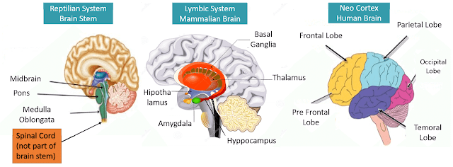
The amygdala is a collection of nuclei found deep within the temporal lobe. The term amygdala comes from Latin and translates to "almond," because one of the most prominent nuclei of the amygdala has an almond-like shape. Although we often refer to it in the singular, there are two amygdalae—one in each cerebral hemisphere.
What is the amygdala and what does it do?
The amygdala is recognized as a component of a group of brain structures referred to collectively as the limbic system, and is thought to play important roles in emotion and behavior. It is best known for its role in the processing of fear, although as we’ll see, this is an oversimplified perspective on amygdala function.
Our modern understanding of amygdala function can be traced back to the 1930s, when Heinrich Kluver and Paul Bucy removed the amygdalae of rhesus monkeys and saw drastic effects on behavior. Among other things, the monkeys became more docile and seemed to display little fear. The constellation of behavior that resulted from amygdalae removal was called Kluver-Bucy syndrome, and it led to the amygdala being investigated for its role in fear.
Since, the amygdala has become best known for its role in fear processing. When we are exposed to a fearful stimulus, information about that stimulus is immediately sent to the amygdala, which can then send signals to areas of the brain like the hypothalamus to trigger a "fight-or-flight" response (e.g., increased heart rate and respiration to prepare for action).
Interestingly, research suggests that information about potentially frightening things in the environment can reach the amygdala before we are even consciously aware that there’s anything to be afraid of. There is a pathway that runs from the thalamus to the amygdala, and sensory information about fearful stimuli may be sent along this pathway to the amygdala before it is consciously processed by the cerebral cortex. This allows for the initiation of a fear reaction before we even have time to think about what it is that’s so frightening.
This type of reflexive response can be useful if we really are in great danger. For example, if you are walking through the grass and a snake darts out at you, you don't want to have to spend a lot of time cognitively assessing the danger the snake might pose. Instead, you want your body to experience immediate fear and jump backward without having to consciously initiate this action. The direct pathway from the thalamus to the amygdala may be one way to achieve this type of response.
Watch this 2-Minute Neuroscience video to learn more about the amygdala.
In addition to its involvement in the initiation of a fear response, the amygdala also seems to be very important in forming memories that are associated with fear-inducing events. For example, if you take mice with intact amygdalae and play a tone right before you give them an uncomfortable foot shock, they will very quickly begin to associate the tone with the unpleasant shock. Thus, they will display a fear reaction (e.g., freezing in place) as soon as the tone is played, but before the shock is initiated. If you attempt this experiment in mice with lesions to the amygdalae, however, they display an impaired ability to "remember" that the tone preceded the foot shock. You can play the tone and they will continue about their business as if they have no bad memories associated with the noise.
It shouldn't be too surprising (given its role in fear processing) that the amygdala might also play a role in anxiety. While fear is considered a response to a threat that is present, anxiety involves the dread that accompanies thinking about a potential threat—one that may or may not ever materialize. A number of studies suggest that the amygdala is involved in experiencing anxiety, and that it may be overactive in people with anxiety disorders. However, as is the case with most human behaviors, anxiety likely involves a network of brain areas, so activity in the amygdala doesn’t tell us all we need to know about the emotion.
Although the amygdala is well-known for its role in fear responses, there is now a great deal of evidence that suggests its contribution to behavior is much more complex. For example, the amygdala seems to be involved with the formation of positive memories, like earning a reward in an experiment. And damage to the amygdala can impair the ability to form these positive memories, just like it can affect the ability to form memories about negative events like the foot shock mentioned above.
Because of research like this, researchers have been forced to expand the role of the amygdala beyond that of just a threat detector/fear generator. One popular perspective suggests that the amygdala is involved with evaluating things in the environment to determine their importance—whether their value is positive or negative—and generating emotional responses to those stimuli that are considered important. It also may be involved in the consolidation of memories that have some strong emotional component, regardless of whether the associated emotions are pleasant or unpleasant. Thus, our understanding of the function of the amygdala is still evolving, and we likely have much more to learn before we can fully catalog the activities of this complex structure.
History
The experiments of Heinrich Kluver and Paul Bucy in the 1930s eventually led to the amygdala being identified as an area of interest in the neuroscience of human behavior. Kluver had been studying (and taking) the psychedelic drug mescaline, and was interested in what part of the brain might be responsible for producing the unique hallucinatory effects of the drug. Kluver hypothesized that the brain region in question might reside in the temporal lobe, because large doses of mescaline given to monkeys could produce side effects that resembled the symptoms of a type of epilepsy called temporal lobe epilepsy.
Kluver recruited a young neurosurgeon named Paul Bucy to help him explore his hypothesis by surgically removing parts of the temporal lobe in monkeys. If the temporal lobe was critical for the effects of mescaline, Kluver hypothesized, then removing enough brain tissue from the region should render the drug ineffective.
The first monkey Kluver and Bucy operated on was an aggressive and unruly monkey named Aurora. Bucy removed most of Aurora's right and left temporal lobes, and afterwards he and Kluver noted some drastic changes to Aurora's personality. Previously, Aurora had been hostile and difficult to control, but now she was placid and easy to work with. In fact, she seemed almost incapable of anger; she also displayed no fear reactions. When Kluver and Bucy published a description of Aurora's changes in personality, it was the first well-known study to link the temporal lobe to emotion. The constellation of behavior that appeared after temporal lobe damage came to be known as Kluver-Bucy syndrome.
A couple of decades later, another scientist named Larry Weiskrantz found that he could elicit Kluver-Bucy syndrome in monkeys just by removing the amgdalae (which are found in the temporal lobes). Weiskrantz's work led other researchers to focus more on the amygdala's role in emotional responses.
While Weiskrantz hypothesized that the amygdala might be involved in enabling monkeys to respond emotionally to both positive and negative stimuli in the environment, many researchers after him focused more on the negative emotions linked to the structure. A large number of studies specifically investigated the amygdala's role in fear.
Often these studies involved what is known as a fear conditioning paradigm. Fear conditioning experiments promote the learning of a fearful response to something that previously didn't inspire any fear. These experiments typically involve taking a subject (such as a rodent) and exposing it to a stimulus (such as a beeping tone) that the animal has no positive or negative experience with. Next, the neutral stimulus is paired with something the animal will definitely perceive as negative (such as a mild electrical shock). If this is done enough times, the rodent will eventually begin to display signs of fear when the tone is played, even when it isn't immediately followed by the shock.
Researchers found that damage to the amygdala disrupts fear conditioning. In other words, if you damage a rat's amygdalae and then put it into a fear conditioning experiment, it won't learn to fear the tone—no matter how many times the tone is paired with a shock. Further research in rodents found that neurons in the amygdala are highly active when rodents hear a tone that has been linked to a fear-inducing stimulus. And studies in humans helped to confirm the role of the amygdala in learning about and experiencing fear-inducing things.
As mentioned above, subsequent research has demonstrated that the amygdala is involved in much more than just fear. For example, more recent experiments have shown that the amygdala also plays a role in learning about positive things (like rewards), and amygdala damage can disrupt the ability to form memories about those positive stimuli. Neuroscientists today generally support a perspective that the amygdala is important in creating emotional responses to both positive and negative things in our environment, as well as in forming memories about those emotionally-salient things.
Disorders involving the amygdala
There are several neurological disorders associated with damage to the amygdala. One, as discussed above, is Kluver-Bucy syndrome. Kluver-Bucy syndrome is rare in humans, but can occur after brain trauma, neurodegenerative disease, or an infection that reaches the brain. The symptoms vary from case to case, but might include placidity, an irresistible urge to put various objects (appropriate and inappropriate) in the mouth (also known as hyperorality), and an uncontrollable appetite.
Urbach-Wiethe disease is a rare genetic disorder that can cause calcification of brain tissue in the temporal lobes; this calcification can cause damage to the amygdalae. While Urbach-Wiethe disease is an exceedingly rare condition, it is thought to be the cause of amygdala damage in one of the best-known medical cases alive today: SM. SM, who is only known by her initials to protect her anonymity, has a well-documented inability to experience fear. Over the past several decades, researchers have put SM into a variety of experimental conditions designed to elicit fear. Only one—forcing her to breathe air that was about 35% carbon dioxide (a solution that causes people to struggle to breathe and often elicits panicked reactions)—led to a fearful reaction from SM. SM has Urbach-Wiethe disease, and it has caused severe damage to her amygdalae. Because of her inability to experience most types of fear coupled with her amygdala damage, SM is commonly used as an demonstration of the important role the amygdala plays in fear.
The amygdala is also thought to be involved in certain types of temporal lobe epilepsy, which might explain some characteristics of temporal lobe seizures, such as feelings of fear and strong emotional memories. Additionally, the amygdala is implicated in some of the cognitive and behavioral symptoms of neurodegenerative dementias, like Alzheimer's disease. Studies suggest, for example, that the death of neurons in the amygdala in Alzheimer's disease may be a substantial contributor to the memory loss characteristic of the condition.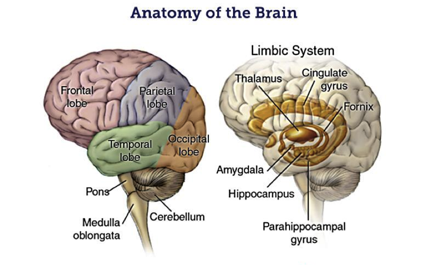
A long list of studies have suggested the involvement of the amygdala in various psychiatric disorders. For example, as mentioned above, increased activity in the amygdala is linked to anxiety, and is thought to be a potential factor in anxiety disorders. Additionally, overactivity in the amygdala is hypothesized to play a role in the symptoms of post-traumatic stress disorder (PTSD). One perspective on the amygdala's contribution to PTSD suggests that the region is hyperactive when patients are exposed to something related to their past trauma (e.g., a photograph). This increased amygdala activity might cause the individual to experience an intense fear reaction, which prompts the brain and body to respond to the stimulus almost as if the trauma is happening all over again. Models of amygdala function in PTSD also often suggest a role for the prefrontal cortex, which typically is thought to inhibit excessive amygdala function when there is not an actual threat in the environment.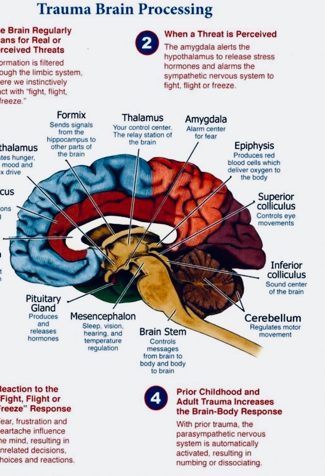 In patients with PTSD, these inhibitory mechanisms may be deficient, causing increased amygdala activity to continue on unabated.
More advanced anatomy
As mentioned above, the name amygdala comes from the Latin word for almond, and the amygdala earned this designation because it is partially composed of an almond-shaped structure found deep within the temporal lobes. The almond-shaped structure, however, is just one nucleus of the amygdala (the basal nucleus)—for although it is often referred to as one entity, the amygdala is actually made up of a collection of nuclei along with some other distinct cell groups. The nuclei of the amygdala include the basal nucleus, accessory basal nucleus, central nucleus, lateral nucleus, medial nucleus, and cortical nucleus. Each of these nuclei can also be partitioned into a collection of subnuclei (e.g., the lateral nucleus can be divided into the dorsal lateral, ventrolateral, and medial lateral nuclei).
Exactly how the amygdala should be divided anatomically has been the subject of some debate, and no clear consensus has been reached.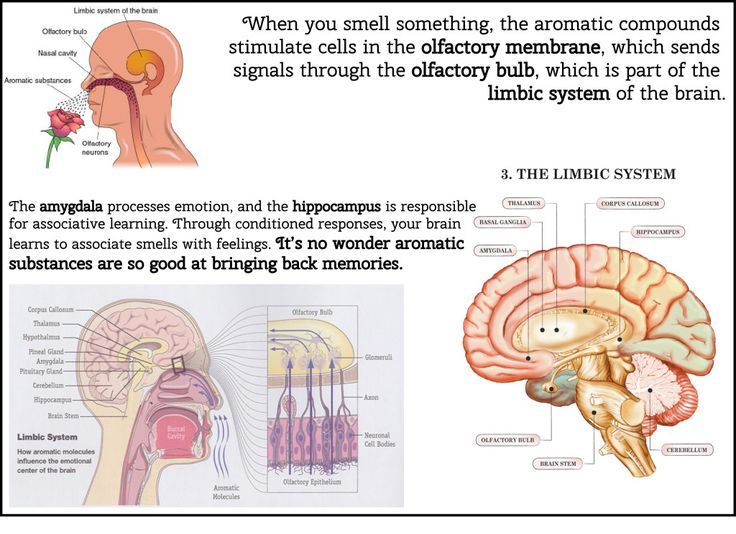 Many researchers group the lateral, basal, and accessory basal nuclei together into a structure referred to as the basolateral complex or basolateral amygdala, and sometimes the cortical and medial nuclei are aggregated as the cortico-medial region. However, there is even a lack of consistency in the application of these terms. For example, some investigators use the basolateral designation to refer to the complex mentioned above, while others use it to refer to just the basal nucleus or basolateral nucleus specifically. Thus, the anatomy of the amygdala is much more complex than is often implied in simple descriptions of the structure. Indeed, the complexity is significant enough that neuroanatomists still have a hard time agreeing on how the different components of the amygdala should be categorized.
In addition to its anatomical diversity, the amygdala has abundant connections throughout the brain—connections that are widespread and divergent enough to suggest many functions beyond just threat detection. For example, many areas of the prefrontal cortex as well as sensory areas throughout the brain have bidirectional connections with the amygdala. The amygdala also has projections that extend to the hippocampi, basal ganglia, basal forebrain, hypothalamus, and a variety of other structures.
References (in addition to linked text above):
Benarroch EE. The amygdala: functional organization and involvement in neurologic disorders. Neurology. 2015 Jan 20;84(3):313-24. doi: 10.1212/WNL.0000000000001171. Epub 2014 Dec 19. PMID: 25527268.
Dingman M. Your Brain, Explained. Boston, MA. Nicholas Brealey Publishing; 2019.
LeDoux J. The amygdala. Curr Biol. 2007 Oct 23;17(20):R868-74.
Learn more:
2-Minute Neuroscience: Amygdala
The amygdala: Beyond fear
Neuroscience for all. Details. The core of fear: what is the amygdala
The amygdala (almond) is a small part of the brain, named for its resemblance to the kernel of an almond.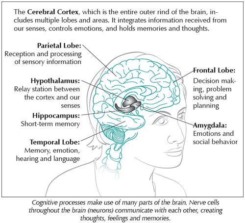 Sometimes in Russian-language literature it is called an amygdala, but this is not quite the correct direct transliteration of the English name. The amygdala is a paired section, the tonsils are located in the temporal lobes of both hemispheres. They belong to the limbic system - an ancient part of the brain that controls autonomic functions, some physiological reactions and emotions. In the formation of the latter, the tonsils are involved. In addition, they are associated with the functioning of memory and decision making. nine0004
Names: Almond -shaped body, tonsil
English name: Amygdala
Latin name: Corpus Amygdaloideum 9000 9000
three groups of nuclei. The basolateral ones are responsible for emotions, the cortical ones are associated with taste sensations, and the medial ones are associated with smell. Their joint work can play a protective function - for example, an unpleasant taste or smell makes a person experience negative emotions and stay away from what causes them - spoiled food that can be poisoned, or waste products that can contain dangerous bacteria. nine0007
The amygdala is shown in purple
In addition, the amygdala is connected to the hippocampus, which is responsible for long-term memory. Therefore, after meeting with something terrible or unpleasant, its image will be fixed in the memory, and subsequently it will be possible to recognize it in time and avoid new contact.
Interestingly, depending on the location, the amygdala generates different emotions. In a study by specialists from the University of Provence, it turned out [1] that electrical stimulation of the right amygdala causes negative emotions - sadness, fear, anxiety. And the stimulation of the left - more often happiness and only sometimes - unpleasant experiences. nine0007
Amygdala nuclei
It is usually said that in men the amygdala is larger than in women, but develops more slowly [2] - the female reaches its peak of development on average 1.5 years earlier. In fact, it is not very clear whether this is so.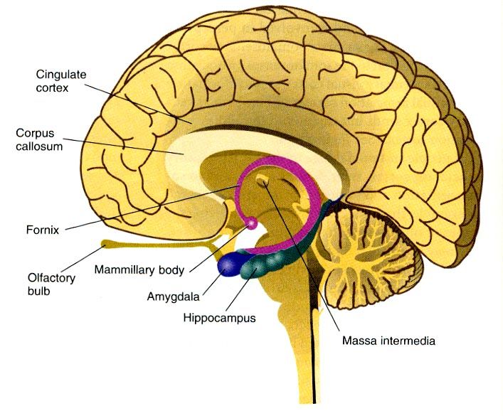
For example, a 2017 meta-analysis [3], based on 46 studies and data from 6726 people, says that yes, in general, the amygdala is on average 0.3 cubic centimeters larger in men than in women. This gives us a 10 percent increase in volume. But if we renormalize it to the fact that the brain itself, on average, in men is 11-12 percent larger than in women (this has nothing to do with the fact that men are smarter - they are just slightly larger than women, and the scatter of the normal volume parameters brain in a person as a whole differs not by percentages, but by several times - from a little more than 1000 cc for Anatole France to a little more than 2000 cc for Ivan Turgenev), it turns out that there is no difference by gender in the relative volume the amygdala is not observed. nine0007
Meta-analysis of 2014 [4], using 126 studies, on the contrary, indicates an increase in the relative volume of the left amygdala in men (including - among other differences, we see the hippocampus and the insula).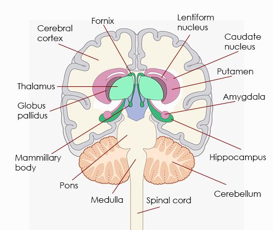
Illustration from [4]. Blue shows areas that are larger in men than in women
In addition, regardless of gender, the left amygdala matures 1.5-2 years faster than the right. The early development of the left amygdala provides the ability to respond to dangers in childhood. nine0007
The size of the amygdala is related to the number of social contacts that a person maintains, the social groups to which he belongs - the larger the amygdala, the more complex the network of social interactions. In particular, the size of the amygdala is associated with the ability to remember the appearance of other people and recognize their emotions.
In Urbach-Wite disease, an extremely rare genetic disease described in 1929 by Erich Urbach and Camillo Witte (about 400 known cases to date), the amygdala can be destroyed. For a long time it was believed that this makes patients completely fearless, but in 2013, American scientists found out that it is still possible to scare such people [5]. This requires inhalation with a high content of carbon dioxide, about 35 percent. This concentration caused not just fear, but a panic attack in three subjects. nine0007
Erich Urbach
The connection between the amygdala and fear suggests that amygdala activity may play a role in the development of anxiety disorders. For example, stimuli reminiscent of an unpleasant experience can cause the amygdala to signal the body to prepare for a fight or run away. Perhaps this is the reason, for example, the mechanism of panic attacks.
This is also evidenced by a study by an international group of scientists, published back in 2011 in the journal Nature [6]. They found that in the amygdala during stress, the accumulation of neuropsin protein is activated. It starts a chain of chemical reactions that lead to an increase in the activity of the amygdala itself. The researchers hypothesized that the activity of the neuropsin signaling pathway somehow "freezes" in people with post-traumatic stress disorder (PTSD), anxiety and panic disorders.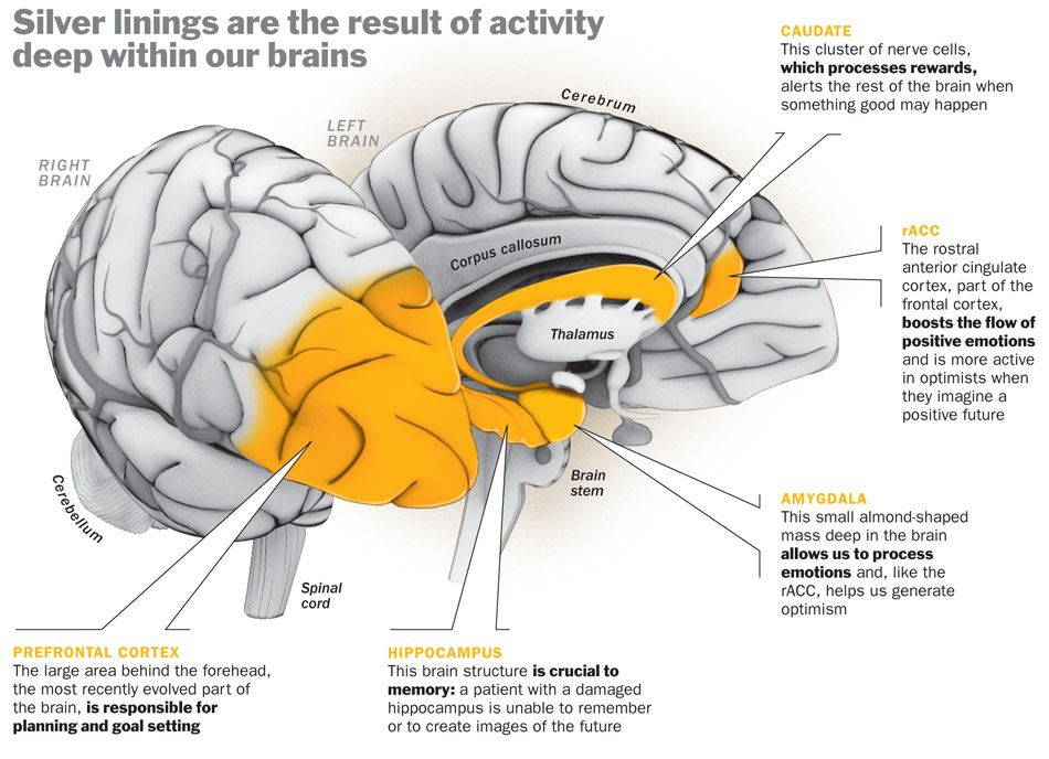 nine0007
In addition, PTSD patients have a surge in amygdala activity when they look at pictures of people who are afraid. Increased activity of the amygdala and in bipolar disorder.
As a last resort for temporal lobe epilepsy, violent outbursts, self-harm and some other "extreme" disorders, amygdalotomy is used - the destruction of the amygdala. This has little effect on memory and intellectual abilities, but affects the recognition of faces and the emotions reflected on them. nine0007
Research on the effects of amygdala removal has been going on since the 19th century. Experiments have shown that such an operation reduces aggressiveness in monkeys (Klüver-Bucy syndrome). In the 20th century, during the heyday of psychosurgery, psychiatrists also took up people. In the vast majority of patients, after destroying the tonsils with a mixture of oil and wax, outbursts of aggression and increased excitability passed. Later, electrodes were used for the operation.
Today, amygdalotomy is rare - the medical community is quite skeptical about such a gross intervention in the brain for the treatment of mental disorders. In addition, the number of pharmaceuticals that help correct the patient's condition has increased. nine0004
Akiko Uematsu, Mie Matsui, Chiaki Tanaka, Tsutomu Takahashi, Kyo Noguchi, Michio Suzuki, Hisao Nishijo PLOS One, Published: October 9, 2012, https://doi.org/10.1371/journal.pone.0046970
7 3. Meta-analysis reveals a lack of sexual dimorphism in human amygdala volume human brain structure
Amber N.V. Ruigrok, Gholamreza Salimi-Khorshidi, Meng-Chuan Lai, Simon Baron-Cohen, Michael V. Lombardo, Roger J. Tait, and John Suckling. Neurosci Biobehav Rev. February 2014; 39(100): 34–50. doi: 10.1016/j.neubiorev.2013.12.004
5. Fear and panic in humans with bilateral amygdala damage
Feinstein, J. S., Buzza, C., Hurlemann, R., Follmer, R. L., Dahdaleh, N. S., Coryell, W. H., ... Wemmie, J. A. (2013). Nature Neuroscience, 16, 270. Retrieved from https://doi.org/10.1038/nn.3323
6. Neuropsin cleaves EphB2 in the amygdala to control anxiety
Attwood, B. K., Bourgognon, J.-M., Patel, S., Mucha, M., Schiavon, E., Skrzypiec, A. E., … Pawlak , R. (2011). Nature, 473(7347), 372–375. https://doi.org/10.1038/nature09938
An area in the brain responsible for our attitude towards animals
under the right temple. As neurophysiologists have established, it is this wise organ that has been responsible for our interaction with animals for millions of years. nine0007
Everyone has a friend or acquaintance who is ready to caress and take care of every homeless animal that catches their eye. There will always be those who would prefer to walk three miles around any object resembling a dog or even a cat, confirming the universal law derived by one Chinese sage that all animals, without exception, viewed from afar, must turn into flies.
But regardless of how you feel about animals in general and individual representatives of the animal kingdom in particular, before reacting to an animal, you must establish the very fact of its presence in the space around you. It turns out that
the basic function that makes it possible to distinguish animals from other objects is performed by a certain area of the human brain amygdala - two small tonsils symmetrically located inside the temporal lobes of both hemispheres.
A group of neurophysiologists from the California Institute of Technology (CalTech) has discovered an "animal plug" in the amygdala, whose article describing this discovery is published in Nature Neuroscience .
The amygdala, which is also called the "emotion processor", is responsible for the formation of such an important emotion for survival as fear, and the subsequent fixation of the emotional response provoked by one or another stimulus (that is, recognized information entering the brain from outside), in " long memory. “Faults” in the formation of negative emotions (anxiety, fear) in the amygdala can provoke such common mental disorders as, for example, panic attacks, and the biochemical mechanism of such disorders associated with excessive accumulation of hormone 9 in the amygdala0145 neuropsin , most recently was finally deciphered by by a group of researchers from the University of Leicester (UK).
The amygdala plays a critical role in regulating basic human emotions, but the CalTech team found that of all the stimuli that trigger this "emotion processor," there is one that irritates the amygdala more than others.
And the animals turned out to be such an irritant.
This was found out in the course of an experiment conducted with the participation of patients of the Medical Center. Ronald Reagan at the University of California, Los Angeles, suffering from severe forms of epilepsy, not amenable to drug correction. Before surgery, electrodes are implanted into the brain of such patients to help doctors localize the problematic epileptic area. Neuroscientists decided to use (with the consent of the patients themselves, of course) this procedure to record the signals produced by neurons located in different areas of the brain that process different visual information. nine0007
41 patients with implanted electrodes participated in the experiment. During each of the 111 sessions, each of them was randomly shown a series of approximately one hundred photographs of various people, buildings, landscapes, objects, rooms and animals.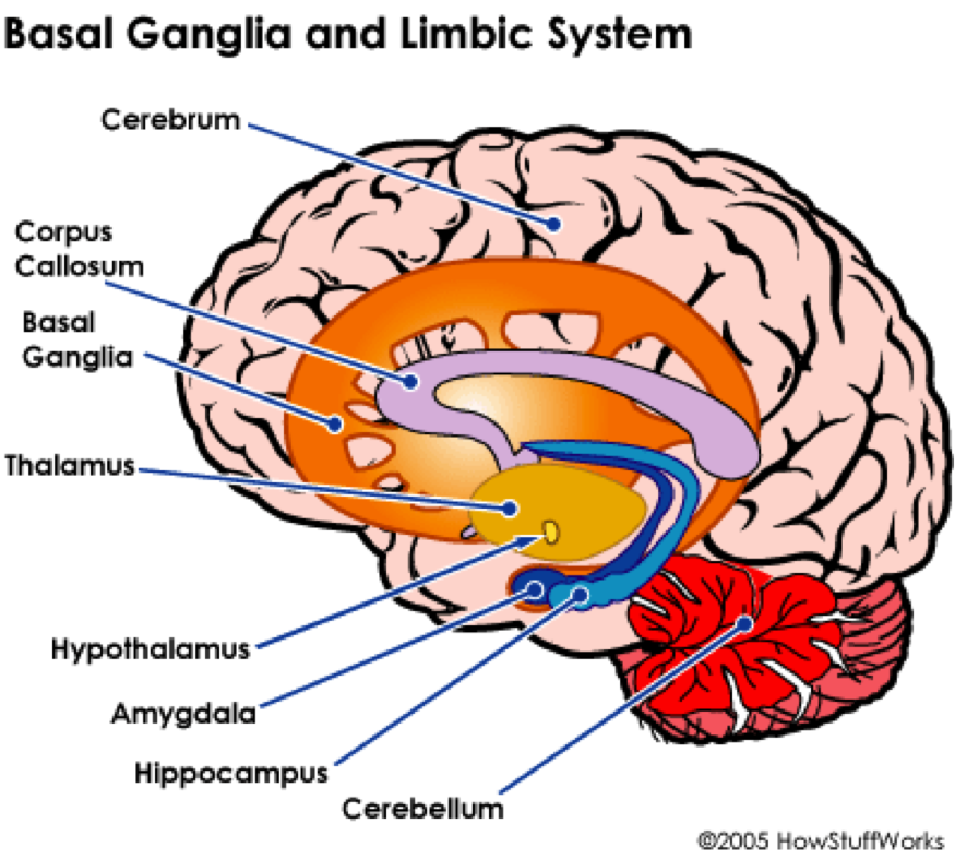 In total, information was collected from 1445 individual neurons located in the amygdala (489), the hippocampus (549) and the entorhinal cortex (407) - the interface connecting the hippocampus with the cerebral cortex and playing an important role in the formation episodic, autobiographical and spatial memory in humans. nine0007
Analysis of the collected data showed that
amygdala neurons fired selectively and rapidly when photographs of animals were shown to patients, and more intensely and with a shorter temporal response than neurons in the hippocampus and entorhinal cortex, which did not demonstrate any thematic specialization (see Fig. schedule).
close
100%
To exclude the "epileptic" theory, the experiment was repeated, but with the participation of healthy people and a magnetic resonance scanner that is not able to take signals from individual neurons (in this sense, the data obtained as a result of direct measurements neuroactivity, more valuable and more accurate), but also recorded an increased amygdala response to images of animals. nine0007
Other possible theories (asymmetric responses in right- and left-handed people, statistical errors, noises spontaneously emitted by neighboring neurons) were also successively ruled out.
“The selective response of animal amygdala neurons did not depend on the emotional content of the photographs, and whether these animals are dangerous to humans or not,” explains Florian Mormann, who led the CalTech experiment and lead author of the article.
"Remarkably, this behavior of neurons was observed only in the amygdala, located in the right hemisphere",
, he notes.
This asymmetry, the authors believe, confirms the theory that in the early stages of vertebrate evolution there was a specialization of the right hemisphere of the brain, "filtering" and processing visual stimuli associated with the activities of other organisms. “The amygdala is an evolutionarily very ancient part of the brain that carries information about huge time spans of our biological history.