Brain waves that occur during sleep are
The Different Kinds of Sleep
Each of us spends about one-third of our life asleep. Or, to put it another way, by the time you’re 75, you will have spent 25 years sleeping. Sleep is part of the life of all higher vertebrates. Suppressing sleep for an extended period has dramatic effects on an organism’s physiological equilibrium. In short, sleeping is almost as important as eating or breathing.
From a behavioral standpoint, sleep is defined by four criteria: reduced motor activity, diminished responses to external stimuli, posture (lying down with eyes closed) and relatively ready reversibility. These criteria distinguish sleep from comas and from hibernation.
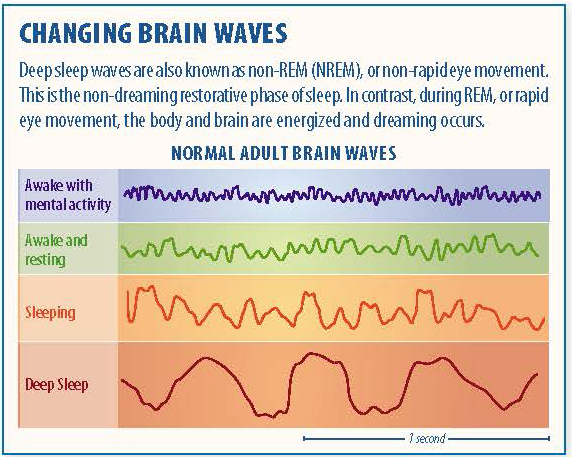
Compared with wakefulness and REM sleep, non-REM sleep is characterized by an electroencephalogram (EEG) in which the waves have greater amplitude and a lower frequency. From the time you fall asleep to the time you reach the deepest non-REM sleep, about 1 ½ hours later, the amplitude of these waves increases continuously, while their frequency diminishes correspondingly.
Scientists have assigned names to four frequency ranges of waves that can be distinguished in an EEG trace. From the highest to the lowest frequency, these waves are as follows.
- Beta waves: have a frequency range from 13-15 to 60 Hz and an amplitude of about 30 µV. Beta waves are the ones registered on an EEG when the subject is awake, alert, and actively processing information. Some scientists distinguish the range above 30-35 Hz as gamma waves, which may be related to consciousness–that is, the making of connections among various parts of the brain in order to form coherent concepts.
- Alpha waves: have a frequency range from eight to twelve Hz and an amplitude of 30 to 50 µV. Alpha waves are typically found in people who are awake but have their eyes closed and are relaxing or meditating.
- Theta waves: have a frequency range from three to eight Hz and an amplitude of 50 to 100 µV. Theta waves are associated with memory, emotions, and activity in the limbic system.

- Delta waves: range from 0.5 to three or four Hz in frequency and 100 to 200 µV in amplitude. Delta waves are observed when individuals are in deep sleep or in a coma.
- Lastly, when there are no brain waves present, the EEG shows a flat-line trace, which is a clinical sign of brain death.
These four types of brain waves, and others discussed below, are important criteria that have been used to define four distinct stages of non-REM sleep. Obviously, falling into a deeper and deeper sleep as the night progresses is actually a gradual, continuous process, but these four stages still provide a convenient means of describing the relative depth of non-REM sleep.
Stage One non-REM sleep begins when you first lie down and close your eyes. After a few sudden, sharp muscle contractions in the legs, the muscles relax. Then, as you continue falling asleep, the rapid beta waves of wakefulness are replaced by the slower alpha waves of someone who is relaxed with their eyes closed.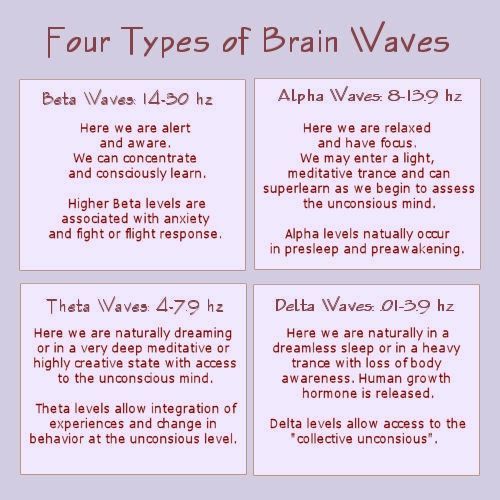 Soon, the even slower theta waves begin to emerge.
Though your reactions to stimuli from the outside world diminish, Stage One is still the phase of sleep from which it is easiest to wake someone up. In experiments where people are awakened from Stage 1 sleep and asked about their state of consciousness, they usually report that they had just fallen asleep or had been in the process of doing so. They also often report having had stray thoughts and short dreams. Each period of Stage One sleep generally lasts three to twelve minutes.
Stage Two non-REM sleep is a stage of light sleep in which the frequency of the EEG trace decreases further while its amplitude increases. The theta waves characteristic of Stage Two sleep are interrupted by occasional series of high-frequency waves known as sleep spindles. These bursts of activity have a frequency of eight to fourteen Hz and an amplitude of 50 to 150 µV. Sleep spindles generally last one to two seconds. They are generated by interactions between thalamic and cortical neurons.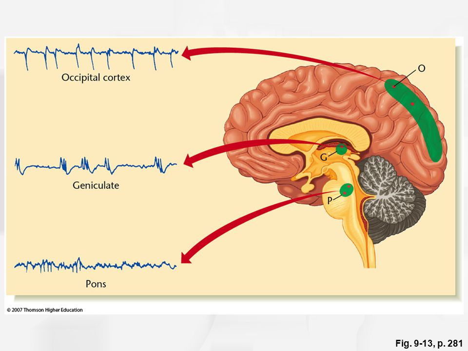
During Stage Two sleep, the EEG trace may also show a fast, high-amplitude wave form called a K-complex. The K-complex seems to be associated with brief awakenings, often in response to external stimuli.
Stage Three non-REM sleep marks the passage from moderately to deep sleep. Delta waves appear and soon account for nearly half of the waves in the EEG trace. Sleep spindles and K-complexes still occur, but less often than in Stage Two. The greater activity observed in the electro-oculogram (EOG) trace during stages three and four reflects the greater amplitude of EEG activity in the prefrontal areas, rather than eye movements.
Stage Three lasts about 10 minutes during the first sleep cycle of the night but accounts for only about 7 percent of a total night’s sleep. During Stage Three, the muscles still have some tone, and sleepers show very little response to external stimuli unless they are very strong or have a special personal meaning (for example, when someone calls your name, or when a baby cries within earshot of its mother).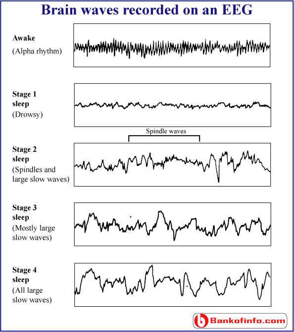
Stage Four non-REM sleep is the deepest, the one in which we sleep the most soundly. The EEG trace is dominated by delta waves and overall neuronal activity is at its lowest. The brain’s temperature is also at its lowest, and breathing, heart rate and blood pressure are all reduced under the influence of the parasympathetic nervous system.
In adults, Stage Four lasts about 35 to 40 minutes during the first sleep cycle of the night; it accounts for 15 to 20 percent of total sleep time in young adults. The muscles still have their tonus, and some movements of the arms, legs and trunk are possible. This is the stage of sleep that accomplishes most of the body’s repair work and from which it is most difficult to wake someone up. This is also the stage of sleep in which children may have episodes of somnambulism (sleepwalking) and night terrors.
Content Provided By
Stages of Sleep | Introduction to Psychology
Learning Objectives
- Differentiate between REM and non-REM sleep
- Describe the stages of sleep
Sleep is not a uniform state of being.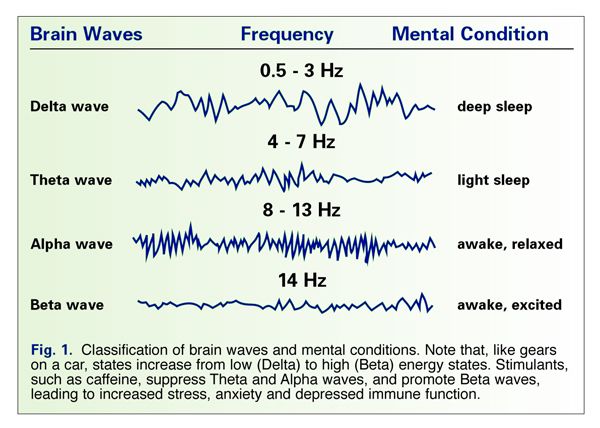 Instead, sleep is composed of several different stages that can be differentiated from one another by the patterns of brain wave activity that occur during each stage. These changes in brain wave activity can be visualized using EEG and are distinguished from one another by both the frequency and amplitude of brain waves. Sleep can be divided into two different general phases: REM sleep and non-REM (NREM) sleep. Rapid eye movement (REM) sleep is characterized by darting movements of the eyes under closed eyelids. Brain waves during REM sleep appear very similar to brain waves during wakefulness. In contrast, non-REM (NREM) sleep is subdivided into three stages distinguished from each other and from wakefulness by characteristic patterns of brain waves. The first three stages of sleep are NREM sleep, while the fourth and final stage of sleep is REM sleep. In this section, we will discuss each of these stages of sleep and their associated patterns of brain wave activity.
[Note that psychologists originally identified four stages of non-REM sleep, but these were revised in 2008, resulting in just three distinct phases of NREM sleep.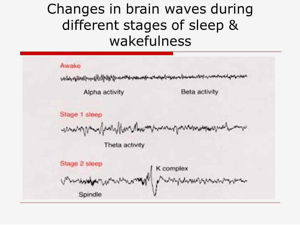 You will see that stage 3 of NREM sleep is sometimes presented as both stage 3 and stage 4 in various texts.]
NREM Stages of Sleep
The first stage of NREM sleep is known as stage 1 sleep. Stage 1 sleep is a transitional phase that occurs between wakefulness and sleep, the period during which we drift off to sleep. During this time, there is a slowdown in both the rates of respiration and heartbeat. In addition, stage 1 sleep involves a marked decrease in both overall muscle tension and core body temperature.
In terms of brain wave activity, stage 1 sleep is associated with both alpha and theta waves. The early portion of stage 1 sleep produces alpha waves, which are relatively low frequency (8–13Hz), high amplitude patterns of electrical activity (waves) that become synchronized. This pattern of brain wave activity resembles that of someone who is very relaxed, yet awake. As an individual continues through stage 1 sleep, there is an increase in theta wave activity.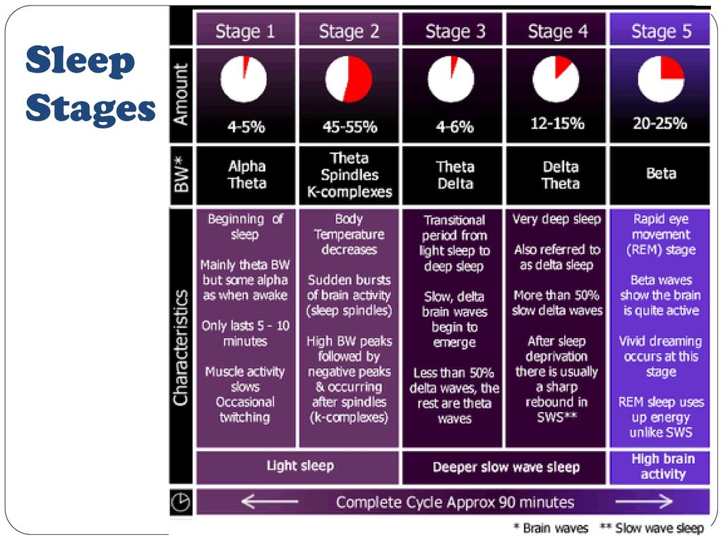 Theta waves are even lower frequency (4–7 Hz), higher amplitude brain waves than alpha waves. It is relatively easy to wake someone from stage 1 sleep; in fact, people often report that they have not been asleep if they are awoken during stage 1 sleep.
Figure 1. Brainwave activity changes dramatically across the different stages of sleep.
As we move into stage 2 sleep, the body goes into a state of deep relaxation. Theta waves still dominate the activity of the brain, but they are interrupted by brief bursts of activity known as sleep spindles (Figure 3). A sleep spindle is a rapid burst of higher frequency brain waves that may be important for learning and memory (Fogel & Smith, 2011; Poe, Walsh, & Bjorness, 2010). In addition, the appearance of K-complexes is often associated with stage 2 sleep. A K-complex is a very high amplitude pattern of brain activity that may in some cases occur in response to environmental stimuli. Thus, K-complexes might serve as a bridge to higher levels of arousal in response to what is going on in our environments (Halász, 1993; Steriade & Amzica, 1998).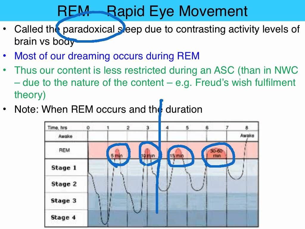
Figure 3. Stage 2 sleep is characterized by the appearance of both sleep spindles and K-complexes.
Stage 3 of sleep is often referred to as deep sleep or slow-wave sleep because these stages are characterized by low frequency (less than 3 Hz), high amplitude delta waves (Figure 4). During this time, an individual’s heart rate and respiration slow dramatically. It is much more difficult to awaken someone from sleep during stage 3 than during earlier stages. Interestingly, individuals who have increased levels of alpha brain wave activity (more often associated with wakefulness and transition into stage 1 sleep) during stage 3 often report that they do not feel refreshed upon waking, regardless of how long they slept (Stone, Taylor, McCrae, Kalsekar, & Lichstein, 2008).
Figure 4. Delta waves, which are low frequency and high amplitude, characterize slow-wave stage 3 sleep.
Try It
REM Sleep
As mentioned earlier, REM sleep is marked by rapid movements of the eyes. The brain waves associated with this stage of sleep are very similar to those observed when a person is awake, as shown in Figure 5, and this is the period of sleep in which dreaming occurs. It is also associated with paralysis of muscle systems in the body with the exception of those that make circulation and respiration possible. Therefore, no movement of voluntary muscles occurs during REM sleep in a normal individual; REM sleep is often referred to as paradoxical sleep because of this combination of high brain activity and lack of muscle tone. Like NREM sleep, REM has been implicated in various aspects of learning and memory (Wagner, Gais, & Born, 2001), although there is disagreement within the scientific community about how important both NREM and REM sleep are for normal learning and memory (Siegel, 2001).
Figure 5. A period of rapid eye movement is marked by the short red line segment. The brain waves associated with REM sleep, outlined in the red box, look very similar to those seen during wakefulness.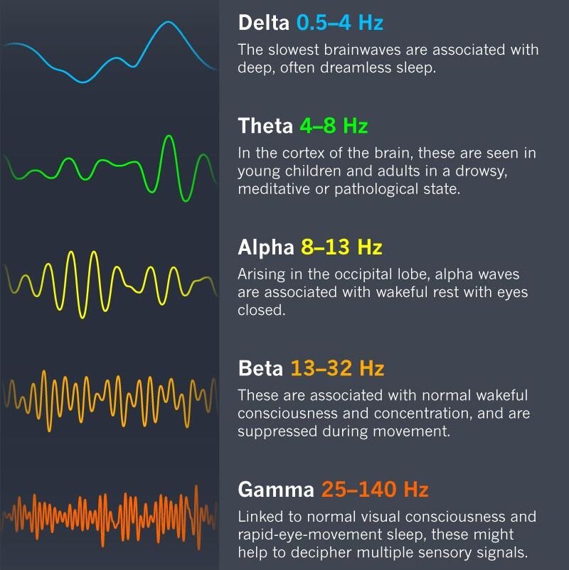
If people are deprived of REM sleep and then allowed to sleep without disturbance, they will spend more time in REM sleep in what would appear to be an effort to recoup the lost time in REM. This is known as the REM rebound, and it suggests that REM sleep is also homeostatically regulated. Aside from the role that REM sleep may play in processes related to learning and memory, REM sleep may also be involved in emotional processing and regulation. In such instances, REM rebound may actually represent an adaptive response to stress in nondepressed individuals by suppressing the emotional salience of aversive events that occurred in wakefulness (Suchecki, Tiba, & Machado, 2012). Sleep deprivation in general is associated with a number of negative consequences (Brown, 2012).
Figure 6. This hypnogram illustrates how an individual moves through the various stages of sleep. Deeper NREM sleep occurs early on in the night, while the duration of REM sleep increases as the night progresses.
Try It
Think It Over
Researchers believe that one important function of sleep is to facilitate learning and memory. How does knowing this help you in your college studies? What changes could you make to your study and sleep habits to maximize your mastery of the material covered in class?
Glossary
alpha wave: type of relatively low frequency, relatively high amplitude brain wave that becomes synchronized; characteristic of the beginning of stage 1 sleep
delta wave: type of low frequency, high amplitude brain wave characteristic of stage 3 sleep
K-complex: very high amplitude pattern of brain activity associated with stage 2 sleep that may occur in response to environmental stimuli
non-REM (NREM): period of sleep outside periods of rapid eye movement (REM) sleep
rapid eye movement (REM) sleep: period of sleep characterized by brain waves very similar to those during wakefulness and by darting movements of the eyes under closed eyelids
sleep spindle: rapid burst of high frequency brain waves during stage 2 sleep that may be important for learning and memory
stage 1 sleep: first stage of sleep; transitional phase that occurs between wakefulness and sleep; the period during which a person drifts off to sleep
stage 2 sleep: second stage of sleep; the body goes into deep relaxation; characterized by the appearance of sleep spindles
stage 3 sleep: third stage of sleep; deep sleep characterized by low frequency, high amplitude delta waves
theta wave: type of low frequency, low amplitude brain wave characteristic of the end of stage 1 sleep
Contribute!
Did you have an idea for improving this content? We’d love your input.
Improve this pageLearn More
Two types of brain waves during sleep determine the process of memorization in rats
Researchers noticed that two types of brain waves fight over whether the rat will remember the new information or forget it. The details of this previously hidden encounter may eventually eventually help explain how some memories are stored in dormant brain, while others are removed, - writes sciencenews.org with referring to Cell .
By distinguishing between these brain waves, new research helps reconcile some seemingly conflicting ideas, including including how memories can be strengthened or weakened during the same stage of sleep. “This will help unify the area of sleep and learning…,” says neuroscientist Gina Po of the California University of Los Angeles, which did not participate in research.
Researchers led by neuroscientist and neurologist Karunesh Ganguly from UC San Francisco teaches rats mechanically control the jet of water only by using their nervous activity.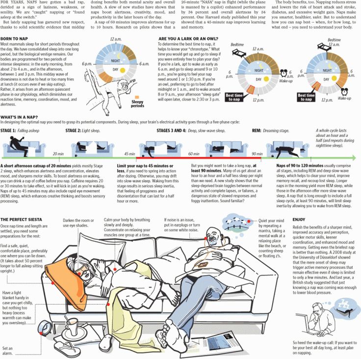 The team soon realized that the success of the rats in learning was highly dependent on whether there was sleep after training.
To study how learning was enhanced during sleep, Ganguly and his team monitored the brains of sleeping rats after they follow the flow of the stream. Scientists have focused on the brain waves that originate in the motor cortex, the part of the brain which controlled the jet of water - during slow sleep. This the sleep stage is usually more than half of an adult's night person.
Two types of brainwaves interested the team. The first, called slow oscillations, and were previously considered important in strengthening memories. Researchers have confirmed this.
The second type, called delta waves, was three to four times more common than slow oscillations, but the role of these waves was a mystery. “We thought that delta waves should do something important because they are so common,” says Ganguly.
The researchers found that stopping the delta waves had the opposite effect of stopping slow oscillations.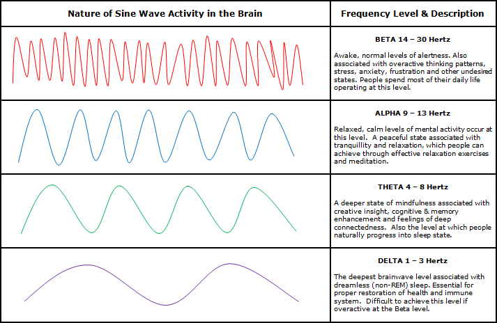 Stop delta waves made the rats perform their task better after how they woke up. According to Poe, “In this study it is quite clear that they are not only different from each other, but also perform diametrically opposed functions,” she says.
Gangly says random slow vibrations help save memories.
It is still unknown how the brain decides which memories keep and which ones to remove. Gangly suspects the key a factor may be the presence of a reward - external, for example, water for rat, or internal, like a pleasant feeling that a person receives from friendly conversation.
The results may ultimately also affect how people will learn to move after strokes. Ganguly says that these people have delta waves more often than usual.
[Photo: sciencenews.org]
Scientists: Sleep detoxifies the brain
Image caption,
Cerebral fluid bathes brain cells continuously, but during sleep it is especially intense
The brain uses periods of sleep to get rid of toxins accumulated in it during the day.
A group of American scientists believe that this mechanism is one of the main causes of sleep.
They found that during sleep, neurons decrease in size and spaces appear between them, which are filled with cerebral fluid.
In an article published in the journal Science, the scientists also suggest that disturbances in the mechanism of removal of toxic proteins may be related to the occurrence of brain diseases.
Biologists have long wondered why all animals go to sleep, despite the fact that it makes them more vulnerable to predators.
It has been known for some time that sleep plays an important role in the formation of memories and the processing of learned information, but scientists at the University of Rochester Medical Center have concluded that one of the main functions of sleep may be to clear the brain.
"The brain has a limited amount of energy, and it looks like it has to choose between two different functional states - awake or sleeping (or clearing)," says Dr.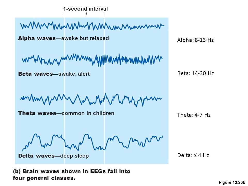 Meiken Nedergaard, one of the researchers.
The results of the scientists' observations are based on the discovery last year of the so-called glymphatic system, which operates in the brain specifically to remove harmful substances.
Scientists who scanned the brains of mice found that during sleep, the glymphatic system increases its activity by 10 times.
Skip the Podcast and continue reading.
Podcast
What was that?
We quickly, simply and clearly explain what happened, why it's important and what's next.
episodes
End of Story Podcast
Brain cells - perhaps the glial cells that surround and support neurons - shrink during sleep. This leads to an increase in the intercellular space in the substance of the brain, which in turn increases the flow of fluid, which removes toxins from the brain.